


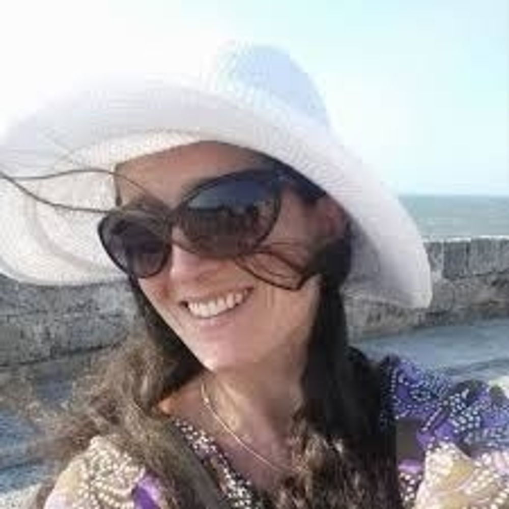
Rachael Ott
@rachaelott.bsky.social
Neurobiologist @UofSC 🔬 Super-resolution #microscopy, adipocytes, cytoskeleton, and #neurodegeneration 🦠
Pinned
Rachael Ott
@rachaelott.bsky.social
· Jan 17

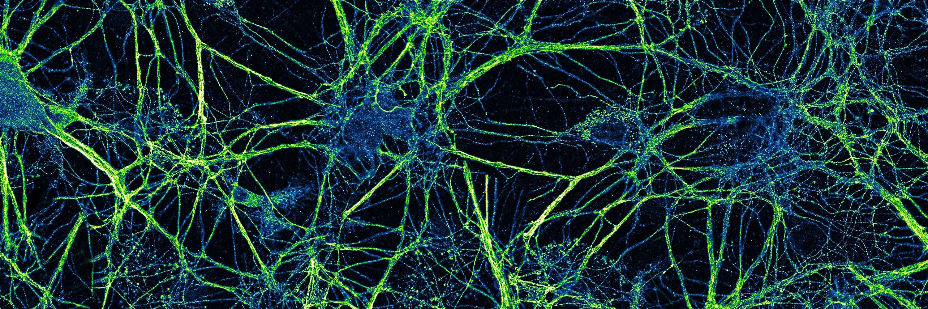
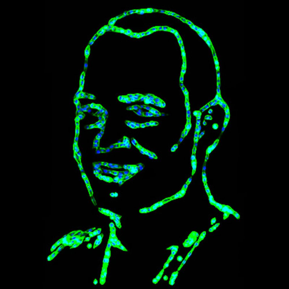
For #FluorescenceFriday, tau (blue) and long loops of neurofilament proteins are shown in a primary hippocampal neuron 🔬 #Neuroscience #Microscopy
Reposted by Rachael Ott

Super proud to be part of SFB1348 — and excited to share that I now officially have my own lab in Münster! 🎉🪰
We’ll study #cellbio & #morphogenesis, focusing on organ sculpting and #chirality in #Drosophila.
I’ll soon hire my first #PhD student — feel free to reach out!
#juniorPI #Science #devbio
We’ll study #cellbio & #morphogenesis, focusing on organ sculpting and #chirality in #Drosophila.
I’ll soon hire my first #PhD student — feel free to reach out!
#juniorPI #Science #devbio

November 25, 2025 at 3:57 PM
Super proud to be part of SFB1348 — and excited to share that I now officially have my own lab in Münster! 🎉🪰
We’ll study #cellbio & #morphogenesis, focusing on organ sculpting and #chirality in #Drosophila.
I’ll soon hire my first #PhD student — feel free to reach out!
#juniorPI #Science #devbio
We’ll study #cellbio & #morphogenesis, focusing on organ sculpting and #chirality in #Drosophila.
I’ll soon hire my first #PhD student — feel free to reach out!
#juniorPI #Science #devbio
Reposted by Rachael Ott
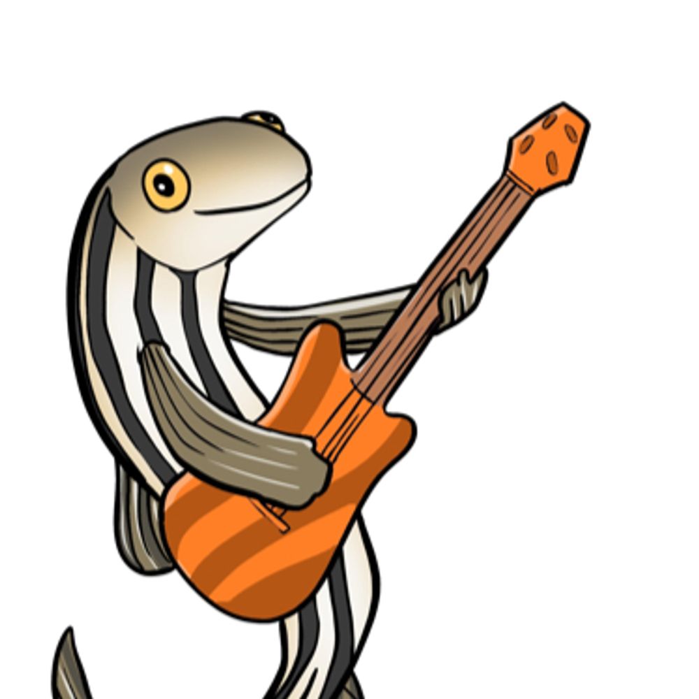
Gemini 3 created this figure in under 30 seconds from a couple reference images and a one sentence prompt
So I’m at a loss for words, honestly #MicroscopyMonday
So I’m at a loss for words, honestly #MicroscopyMonday

November 24, 2025 at 7:11 PM
Gemini 3 created this figure in under 30 seconds from a couple reference images and a one sentence prompt
So I’m at a loss for words, honestly #MicroscopyMonday
So I’m at a loss for words, honestly #MicroscopyMonday
Reposted by Rachael Ott
I like this movie, but a friend of mine likes to complain about the obvious stitching artifacts. I'll try harder next time, Michael. Vimentin (orange) and ER (blue) in an overnight acquisition.
November 25, 2025 at 6:06 AM
I like this movie, but a friend of mine likes to complain about the obvious stitching artifacts. I'll try harder next time, Michael. Vimentin (orange) and ER (blue) in an overnight acquisition.
Reposted by Rachael Ott

Some cells are just in it for the drama!
The bottom 5 microns of cells videoed through a microscope by @EmmaKoory. The middle cell rounds up (for fun?) and subsequently rounds up to divide. We missed so much of the action by just sampling the bottom. @CellBiology
The bottom 5 microns of cells videoed through a microscope by @EmmaKoory. The middle cell rounds up (for fun?) and subsequently rounds up to divide. We missed so much of the action by just sampling the bottom. @CellBiology
November 24, 2025 at 9:19 PM
Some cells are just in it for the drama!
The bottom 5 microns of cells videoed through a microscope by @EmmaKoory. The middle cell rounds up (for fun?) and subsequently rounds up to divide. We missed so much of the action by just sampling the bottom. @CellBiology
The bottom 5 microns of cells videoed through a microscope by @EmmaKoory. The middle cell rounds up (for fun?) and subsequently rounds up to divide. We missed so much of the action by just sampling the bottom. @CellBiology
Reposted by Rachael Ott

Entire creation of new data that has no bearing upon any histological relevance. Remember LLM models are not open and we have no understanding of what NeuN, GFAP, or DAPI means in biological space within the LLM.
This is nonsense. But it looks at first blush as "real" to the uninformed.
Scary.
This is nonsense. But it looks at first blush as "real" to the uninformed.
Scary.
Gemini 3 created this figure in under 30 seconds from a couple reference images and a one sentence prompt
So I’m at a loss for words, honestly #MicroscopyMonday
So I’m at a loss for words, honestly #MicroscopyMonday
November 24, 2025 at 7:51 PM
Entire creation of new data that has no bearing upon any histological relevance. Remember LLM models are not open and we have no understanding of what NeuN, GFAP, or DAPI means in biological space within the LLM.
This is nonsense. But it looks at first blush as "real" to the uninformed.
Scary.
This is nonsense. But it looks at first blush as "real" to the uninformed.
Scary.
Gemini 3 created this figure in under 30 seconds from a couple reference images and a one sentence prompt
So I’m at a loss for words, honestly #MicroscopyMonday
So I’m at a loss for words, honestly #MicroscopyMonday
November 24, 2025 at 7:11 PM
Gemini 3 created this figure in under 30 seconds from a couple reference images and a one sentence prompt
So I’m at a loss for words, honestly #MicroscopyMonday
So I’m at a loss for words, honestly #MicroscopyMonday
Reposted by Rachael Ott

It’s time to talk about epithelial geometry and cancer. How can the architecture of an epithelium affect how tumors will grow and spread? In this thread, @jorgealmagro.bsky.social
November 23, 2025 at 8:01 AM
It’s time to talk about epithelial geometry and cancer. How can the architecture of an epithelium affect how tumors will grow and spread? In this thread, @jorgealmagro.bsky.social
Reposted by Rachael Ott
Still posting cytoskeleton videos, it seems. Actin this time.
Sample: Lifeact-eGFP in HeLa cells.
Modality: Airyscan confocal
Timestamp is mm:ss and the scale bar is 5 µm.
Sample: Lifeact-eGFP in HeLa cells.
Modality: Airyscan confocal
Timestamp is mm:ss and the scale bar is 5 µm.
November 23, 2025 at 3:21 AM
Still posting cytoskeleton videos, it seems. Actin this time.
Sample: Lifeact-eGFP in HeLa cells.
Modality: Airyscan confocal
Timestamp is mm:ss and the scale bar is 5 µm.
Sample: Lifeact-eGFP in HeLa cells.
Modality: Airyscan confocal
Timestamp is mm:ss and the scale bar is 5 µm.
Reposted by Rachael Ott
Happy #FluorescenceFriday! LSCM images of mixed species of Radiolaria, zooplankton. Huygens deconvolved to lure out elusive weak autofluorescence signal and noisy background. Thank you Outi Paloheimo (Imaging Facility of Tampere University)!
Interested in participating? Join: svi.nl/ImageContest
Interested in participating? Join: svi.nl/ImageContest

November 21, 2025 at 8:48 AM
Happy #FluorescenceFriday! LSCM images of mixed species of Radiolaria, zooplankton. Huygens deconvolved to lure out elusive weak autofluorescence signal and noisy background. Thank you Outi Paloheimo (Imaging Facility of Tampere University)!
Interested in participating? Join: svi.nl/ImageContest
Interested in participating? Join: svi.nl/ImageContest
Reposted by Rachael Ott
vimeo.com/1139537563?s...
I uploaded a version of that vimentin IF movie to my vimeo page. It's still compressed, but looks a lot better. Best results when you set the quality to 4k.
I uploaded a version of that vimentin IF movie to my vimeo page. It's still compressed, but looks a lot better. Best results when you set the quality to 4k.

24FPS_VimentinCos7GIF_4320p_ultraHQ
This is "24FPS_VimentinCos7GIF_4320p_ultraHQ" by Andy Moore on Vimeo, the home for high quality videos and the people who love them.
vimeo.com
November 22, 2025 at 4:25 AM
vimeo.com/1139537563?s...
I uploaded a version of that vimentin IF movie to my vimeo page. It's still compressed, but looks a lot better. Best results when you set the quality to 4k.
I uploaded a version of that vimentin IF movie to my vimeo page. It's still compressed, but looks a lot better. Best results when you set the quality to 4k.
Reposted by Rachael Ott

Finally, the first public release of #BigVolumeBrowser, so after teasers, you can try it yourself. For details, please check the announcement post (1/2)
forum.image.sc/t/bigvolumeb...
forum.image.sc/t/bigvolumeb...

BigVolumeBrowser: a new 3D multi volume/mesh/point clould (SMLM) data viewer
Hello everyone, I’d like to share with you another 3D viewer for FIJI, BigVolumeBrowser (full documentation link). It‘s a first initial public release, so there is still space for improvements. Le...
forum.image.sc
November 21, 2025 at 9:29 AM
Finally, the first public release of #BigVolumeBrowser, so after teasers, you can try it yourself. For details, please check the announcement post (1/2)
forum.image.sc/t/bigvolumeb...
forum.image.sc/t/bigvolumeb...
Reposted by Rachael Ott
The law of the jungle.
Interactions of cells in a collective lead to global rotation.
In 80% of the case HUVEC cells turn clockwise.
How many cells does it take for this to happen?
Interactions of cells in a collective lead to global rotation.
In 80% of the case HUVEC cells turn clockwise.
How many cells does it take for this to happen?
November 21, 2025 at 1:07 PM
The law of the jungle.
Interactions of cells in a collective lead to global rotation.
In 80% of the case HUVEC cells turn clockwise.
How many cells does it take for this to happen?
Interactions of cells in a collective lead to global rotation.
In 80% of the case HUVEC cells turn clockwise.
How many cells does it take for this to happen?
Reposted by Rachael Ott
Attempt number seven at uploading this video of intermediate filaments in an enormous COS7 cell. I have a feeling the BlueSky compression will not do it any favors.
November 21, 2025 at 3:54 AM
Attempt number seven at uploading this video of intermediate filaments in an enormous COS7 cell. I have a feeling the BlueSky compression will not do it any favors.
Reposted by Rachael Ott

Cool, the HAK-actin preprint was showcased by @prelights.bsky.social
A good reason to read it if you haven't yet 😜
A good reason to read it if you haven't yet 😜

Reliable actin labelling in expansion #microscopy 🔬
Beautiful #preprint from Olivier Mercey, @lreymond.bsky.social & team @dudinlab.bsky.social @centriolelab.bsky.social
Read the latest #preLight from @kanishka03.bsky.social ⬇️👀
prelights.biologists.com/highlights/h...
Beautiful #preprint from Olivier Mercey, @lreymond.bsky.social & team @dudinlab.bsky.social @centriolelab.bsky.social
Read the latest #preLight from @kanishka03.bsky.social ⬇️👀
prelights.biologists.com/highlights/h...

HAK-actin, U-ExM-compatible probe to image the actin cytoskeleton - preLights
HAK-actin is a new probe that may help researchers see the actin cytoskeleton in sharper detail using expansion microscopy.
prelights.biologists.com
November 19, 2025 at 12:28 PM
Cool, the HAK-actin preprint was showcased by @prelights.bsky.social
A good reason to read it if you haven't yet 😜
A good reason to read it if you haven't yet 😜
Reposted by Rachael Ott
#ZebrafishZunday: Cytoplasmic (or ooplasmic) streaming leads to the segregation of embryo from yolk granules. Credit to @shamipourshayan.bsky.social & @heisenbergcplab.bsky.social. 🧪
November 16, 2025 at 9:31 AM
#ZebrafishZunday: Cytoplasmic (or ooplasmic) streaming leads to the segregation of embryo from yolk granules. Credit to @shamipourshayan.bsky.social & @heisenbergcplab.bsky.social. 🧪
Reposted by Rachael Ott
Lung morphogenesis🦎 Squamate/Lizards
Pressure-driven pushing of 🫁epithelium +
Proliferation-driven expansion of elongating epithelial subbronchi/diverticulae
Smooth muscle mesh provides structural guide for epithelial protrusions
#DevDyn 2025
anatomypubs.onlinelibrary.wiley.com/doi/10.1002/...
Pressure-driven pushing of 🫁epithelium +
Proliferation-driven expansion of elongating epithelial subbronchi/diverticulae
Smooth muscle mesh provides structural guide for epithelial protrusions
#DevDyn 2025
anatomypubs.onlinelibrary.wiley.com/doi/10.1002/...

November 17, 2025 at 12:51 PM
Lung morphogenesis🦎 Squamate/Lizards
Pressure-driven pushing of 🫁epithelium +
Proliferation-driven expansion of elongating epithelial subbronchi/diverticulae
Smooth muscle mesh provides structural guide for epithelial protrusions
#DevDyn 2025
anatomypubs.onlinelibrary.wiley.com/doi/10.1002/...
Pressure-driven pushing of 🫁epithelium +
Proliferation-driven expansion of elongating epithelial subbronchi/diverticulae
Smooth muscle mesh provides structural guide for epithelial protrusions
#DevDyn 2025
anatomypubs.onlinelibrary.wiley.com/doi/10.1002/...
Reposted by Rachael Ott

Check out our recent preprint, "Live longitudinal imaging of meningeal cerebrovascular injury and its sequelae in adult zebrafish."
lnkd.in/gN7z6th4
lnkd.in/gN7z6th4
November 18, 2025 at 2:42 AM
Check out our recent preprint, "Live longitudinal imaging of meningeal cerebrovascular injury and its sequelae in adult zebrafish."
lnkd.in/gN7z6th4
lnkd.in/gN7z6th4
Reposted by Rachael Ott
Meningeal vascular remodeling in 2~4 mo ovariectomized🐭
⏬blood vessel length
⏫blood vessel tortuosity
⏫PDPL+ lymphatic vessel area fraction
Ectopic vessel muscularization
Perivascular scattered αSMA+ cells in dura mater
#Cells_MDPI 2025
www.mdpi.com/2073-4409/14...
⏬blood vessel length
⏫blood vessel tortuosity
⏫PDPL+ lymphatic vessel area fraction
Ectopic vessel muscularization
Perivascular scattered αSMA+ cells in dura mater
#Cells_MDPI 2025
www.mdpi.com/2073-4409/14...

November 15, 2025 at 11:20 PM
Meningeal vascular remodeling in 2~4 mo ovariectomized🐭
⏬blood vessel length
⏫blood vessel tortuosity
⏫PDPL+ lymphatic vessel area fraction
Ectopic vessel muscularization
Perivascular scattered αSMA+ cells in dura mater
#Cells_MDPI 2025
www.mdpi.com/2073-4409/14...
⏬blood vessel length
⏫blood vessel tortuosity
⏫PDPL+ lymphatic vessel area fraction
Ectopic vessel muscularization
Perivascular scattered αSMA+ cells in dura mater
#Cells_MDPI 2025
www.mdpi.com/2073-4409/14...
Reposted by Rachael Ott

During my postdoc, I looked at hundreds of images like this 🤩
These are two apical caulonemal cells from moss Physcomitrium patens stained with MDY64 (shown in shades of orange). The natural autofluorescence of chlorophyll is in cyan.
#microscopymonday #moss #plantcells #plantmicroscopy
These are two apical caulonemal cells from moss Physcomitrium patens stained with MDY64 (shown in shades of orange). The natural autofluorescence of chlorophyll is in cyan.
#microscopymonday #moss #plantcells #plantmicroscopy

November 10, 2025 at 3:33 PM
During my postdoc, I looked at hundreds of images like this 🤩
These are two apical caulonemal cells from moss Physcomitrium patens stained with MDY64 (shown in shades of orange). The natural autofluorescence of chlorophyll is in cyan.
#microscopymonday #moss #plantcells #plantmicroscopy
These are two apical caulonemal cells from moss Physcomitrium patens stained with MDY64 (shown in shades of orange). The natural autofluorescence of chlorophyll is in cyan.
#microscopymonday #moss #plantcells #plantmicroscopy
Reposted by Rachael Ott

Hydra (Hydra vulgaris) ✨Small but mighty! Hydra can regenerate its entire body, even its head 🧠 A classic model for regeneration, stem cell dynamics, and body axis patterning 📸 Image by Daniel Bressan de Andrade #ModelMonday #DevBio #Regeneration

November 11, 2025 at 2:28 AM
Hydra (Hydra vulgaris) ✨Small but mighty! Hydra can regenerate its entire body, even its head 🧠 A classic model for regeneration, stem cell dynamics, and body axis patterning 📸 Image by Daniel Bressan de Andrade #ModelMonday #DevBio #Regeneration
Reposted by Rachael Ott
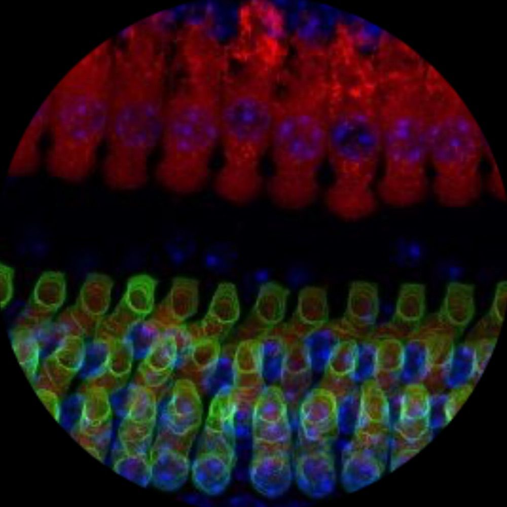
Happy #MicroscopyMonday everyone! 🌟🔬 Here’s an image from Ivy of outer hair cells (OHCs) and inner hair cells (IHCs) from a P20 mouse.. 🐁✨#Microscopy #HearingResearch #Cochlea #Neuroscience #Neuroskyence

November 10, 2025 at 8:01 AM
Happy #MicroscopyMonday everyone! 🌟🔬 Here’s an image from Ivy of outer hair cells (OHCs) and inner hair cells (IHCs) from a P20 mouse.. 🐁✨#Microscopy #HearingResearch #Cochlea #Neuroscience #Neuroskyence
Reposted by Rachael Ott
Check out this sneaky lamin A knockdown cell (pink dot) swimming against the tide (migration into gap) & slithering between its sisters.
It also slides over another nucleus that deforms underneath it.
MCF10A cells, #Spirochrome SPY650-DNA, 20 min time-lapse, taken on a #Zeiss #CellDiscoverer7.
It also slides over another nucleus that deforms underneath it.
MCF10A cells, #Spirochrome SPY650-DNA, 20 min time-lapse, taken on a #Zeiss #CellDiscoverer7.
November 7, 2025 at 4:29 PM
Check out this sneaky lamin A knockdown cell (pink dot) swimming against the tide (migration into gap) & slithering between its sisters.
It also slides over another nucleus that deforms underneath it.
MCF10A cells, #Spirochrome SPY650-DNA, 20 min time-lapse, taken on a #Zeiss #CellDiscoverer7.
It also slides over another nucleus that deforms underneath it.
MCF10A cells, #Spirochrome SPY650-DNA, 20 min time-lapse, taken on a #Zeiss #CellDiscoverer7.
Reposted by Rachael Ott
my first #FluorescenceFriday post! Mouse hippocampus showing microglia, a lysosomal marker (CD68), and Aggrecan, a perineuronal net component. 😍 Image by Dr. Julia Dziabis!

November 7, 2025 at 6:55 PM
my first #FluorescenceFriday post! Mouse hippocampus showing microglia, a lysosomal marker (CD68), and Aggrecan, a perineuronal net component. 😍 Image by Dr. Julia Dziabis!
Reposted by Rachael Ott

Happy #FluorescenceFriday from these primary astrocytes grown on glass slides and robotically stained on the Biocare Oncore Pro X! 🧠🔬🧪
Cyan = GFAP
Royal Purple = ALDH1L1
Salmon = Nuclear Counterstain
Cyan = GFAP
Royal Purple = ALDH1L1
Salmon = Nuclear Counterstain

November 7, 2025 at 7:26 PM
Happy #FluorescenceFriday from these primary astrocytes grown on glass slides and robotically stained on the Biocare Oncore Pro X! 🧠🔬🧪
Cyan = GFAP
Royal Purple = ALDH1L1
Salmon = Nuclear Counterstain
Cyan = GFAP
Royal Purple = ALDH1L1
Salmon = Nuclear Counterstain
Reposted by Rachael Ott

For #FluorescenceFriday - one of my favorites from the archives of a neuron ⚪ and astrocyte 🟢 co-culture. Image credit to Stephanie Page and former research assistant Beth Pattie 🧠🔬🧪

November 7, 2025 at 12:37 PM
For #FluorescenceFriday - one of my favorites from the archives of a neuron ⚪ and astrocyte 🟢 co-culture. Image credit to Stephanie Page and former research assistant Beth Pattie 🧠🔬🧪