


Matt Campbell
@mjcampbell.bsky.social
Pediatric cardiologist. Opinions are my own, not representative of employer and not medical advice. #medsky #echosky #cardiosky
I don't care what anyone said that was an intentional drilling of the batter #worldseries
November 2, 2025 at 1:27 AM
I don't care what anyone said that was an intentional drilling of the batter #worldseries
Reposted by Matt Campbell

In honor of #Halloween, follow along our #SpookyEcho thread! 👻
This is an inverted image of a 3D en face view of the atrial surface of the mitral valve showing a partially dehisced posterior annuloplasty band ("monster's" upper lip and "fangs") & 2 paravalvular defects (eyes).
This is an inverted image of a 3D en face view of the atrial surface of the mitral valve showing a partially dehisced posterior annuloplasty band ("monster's" upper lip and "fangs") & 2 paravalvular defects (eyes).

October 31, 2025 at 1:13 PM
In honor of #Halloween, follow along our #SpookyEcho thread! 👻
This is an inverted image of a 3D en face view of the atrial surface of the mitral valve showing a partially dehisced posterior annuloplasty band ("monster's" upper lip and "fangs") & 2 paravalvular defects (eyes).
This is an inverted image of a 3D en face view of the atrial surface of the mitral valve showing a partially dehisced posterior annuloplasty band ("monster's" upper lip and "fangs") & 2 paravalvular defects (eyes).
Reposted by Matt Campbell

if you live in philly, have a giants jersey, and played corner in high school, please report to the linc immediately ur gonna be guarding devonta smith
October 26, 2025 at 7:03 PM
if you live in philly, have a giants jersey, and played corner in high school, please report to the linc immediately ur gonna be guarding devonta smith
Reposted by Matt Campbell

An oldie, but a goodie.

October 24, 2025 at 6:47 PM
An oldie, but a goodie.
Reposted by Matt Campbell

After a week of ridiculous Republican smears and Trump claiming that “very few people are going to be there,” you just made history. #NoKings

October 18, 2025 at 9:57 PM
After a week of ridiculous Republican smears and Trump claiming that “very few people are going to be there,” you just made history. #NoKings
Reposted by Matt Campbell

No Kings-Houston, TX

October 18, 2025 at 7:58 PM
No Kings-Houston, TX
Reposted by Matt Campbell
Truth! The collective noun for frogs is army. Someone should tell ill-tempered Jesse Watters, who described today’s protest as Dems “trying to do their little Tea Party thing,” that the overseas No Kings protests alone could far outnumber his vaunted series of Tea Party protests. In one day.

October 18, 2025 at 4:52 PM
Truth! The collective noun for frogs is army. Someone should tell ill-tempered Jesse Watters, who described today’s protest as Dems “trying to do their little Tea Party thing,” that the overseas No Kings protests alone could far outnumber his vaunted series of Tea Party protests. In one day.
Reposted by Matt Campbell

The Trump administration has promoted the anti-vax, anti-science movement which has now produced an epidemic www.nbcnews.com/health/healt...

Hundreds of U.S. students quarantined amid measles outbreaks
At least 270 unvaccinated kids are staying home from school as measles continues to spread nationwide. "Expect more," one expert said.
www.nbcnews.com
October 12, 2025 at 11:13 AM
The Trump administration has promoted the anti-vax, anti-science movement which has now produced an epidemic www.nbcnews.com/health/healt...
Reposted by Matt Campbell

Portland frog costume will win Halloween this year. You heard it here first

October 10, 2025 at 10:38 AM
Portland frog costume will win Halloween this year. You heard it here first
Reposted by Matt Campbell
Why in the name of god would you throw home? Why?
October 10, 2025 at 1:40 AM
Why in the name of god would you throw home? Why?
Reposted by Matt Campbell
Went over to the Phillies instagram page to see what’s been going on and was greeted by these two images at the top of their feed

October 9, 2025 at 3:54 AM
Went over to the Phillies instagram page to see what’s been going on and was greeted by these two images at the top of their feed
Reposted by Matt Campbell

Roy Halladay's sons, Braden and Ryan, are throwing out the first pitch to Carlos Ruiz tonight. It's the 15th anniversary of Halladay's no-hitter.
October 6, 2025 at 9:57 PM
Roy Halladay's sons, Braden and Ryan, are throwing out the first pitch to Carlos Ruiz tonight. It's the 15th anniversary of Halladay's no-hitter.
Reposted by Matt Campbell

There's a really nice new study using phone data from Americans who relocated from one city to another to look at the effect of walkable environments on physical activity. E.g., people move TO New York and walk more, they move FROM New York and walk less www.nature.com/articles/s41...

Countrywide natural experiment links built environment to physical activity - Nature
By analysing the smartphone data of 2,112,288 participants, in particular observing and comparing the activity of the same individual in two different environments, we find that increases in the walkability of environments result in increases in daily physical activity, providing evidence of the importance of the built environment on physical health.
www.nature.com
September 29, 2025 at 7:41 AM
There's a really nice new study using phone data from Americans who relocated from one city to another to look at the effect of walkable environments on physical activity. E.g., people move TO New York and walk more, they move FROM New York and walk less www.nature.com/articles/s41...
Reposted by Matt Campbell
Today is #WorldHeartDay! 🫀
In honor of this meaningful day, Elsevier, the publisher of CASE and JASE, is sharing an article from each journal to help spread awareness and important cardiovascular research.
CASE: bit.ly/46ZypHQ
JASE: bit.ly/4pCTlvE
In honor of this meaningful day, Elsevier, the publisher of CASE and JASE, is sharing an article from each journal to help spread awareness and important cardiovascular research.
CASE: bit.ly/46ZypHQ
JASE: bit.ly/4pCTlvE
September 29, 2025 at 2:58 PM
Today is #WorldHeartDay! 🫀
In honor of this meaningful day, Elsevier, the publisher of CASE and JASE, is sharing an article from each journal to help spread awareness and important cardiovascular research.
CASE: bit.ly/46ZypHQ
JASE: bit.ly/4pCTlvE
In honor of this meaningful day, Elsevier, the publisher of CASE and JASE, is sharing an article from each journal to help spread awareness and important cardiovascular research.
CASE: bit.ly/46ZypHQ
JASE: bit.ly/4pCTlvE
What is the correct term for a fake tush push? A butt fake? #flyeaglesfly
September 28, 2025 at 7:21 PM
What is the correct term for a fake tush push? A butt fake? #flyeaglesfly

Reposted by Matt Campbell

#Cardiosky. Blood speckle tracking. No longer only visualisation. Validation of quantitative intraventricular pressure gradient measurement. Congratulation. academic.oup.com/ehjcimaging/...
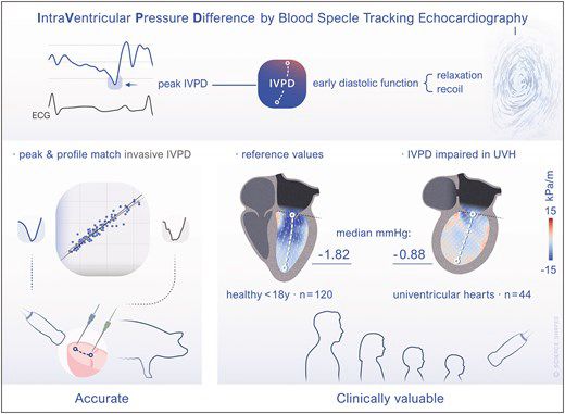
Intraventricular pressure difference estimation based on blood speckle tracking—invasive validation and early clinical application
AbstractAims. Ventricular relaxation creates an intraventricular pressure difference (IVPD) and resultant diastolic suction. Non-invasive estimation by ech
academic.oup.com
September 25, 2025 at 9:55 PM
#Cardiosky. Blood speckle tracking. No longer only visualisation. Validation of quantitative intraventricular pressure gradient measurement. Congratulation. academic.oup.com/ehjcimaging/...
Reposted by Matt Campbell

Novel surgical procedure increases positive outcomes in infants with serious heart condition. @bcmhouston.bsky.social #TexasChildrens www.ahajournals.org/doi/10.1161/...

Technical Advances and Outcomes of Fetal Atrial Septal Intervention for Restrictive or Intact Atrial Septum | Circulation: Cardiovascular Interventions
BACKGROUND: Infants with hypoplastic left heart syndrome with severely restrictive or intact atrial
septum (R/IAS) have low survival. In-utero creation of an atrial septal communication
has been repor...
www.ahajournals.org
September 23, 2025 at 8:36 PM
Novel surgical procedure increases positive outcomes in infants with serious heart condition. @bcmhouston.bsky.social #TexasChildrens www.ahajournals.org/doi/10.1161/...
www.sciencedirect.com/science/arti...
Our cool new article about descending aorta position impact on symptom development in vascular ring patients, can be reliably detected in fetal echo. congrats to Drs. Ostler and Doan especially. #cardiosky #echosky
Our cool new article about descending aorta position impact on symptom development in vascular ring patients, can be reliably detected in fetal echo. congrats to Drs. Ostler and Doan especially. #cardiosky #echosky

Prenatal Prediction of Symptoms in Fetuses With Vascular Ring: A Novel Echocardiographic Marker
www.sciencedirect.com
September 17, 2025 at 1:28 AM
www.sciencedirect.com/science/arti...
Our cool new article about descending aorta position impact on symptom development in vascular ring patients, can be reliably detected in fetal echo. congrats to Drs. Ostler and Doan especially. #cardiosky #echosky
Our cool new article about descending aorta position impact on symptom development in vascular ring patients, can be reliably detected in fetal echo. congrats to Drs. Ostler and Doan especially. #cardiosky #echosky
Backyard villain found on the lemon tree today

September 16, 2025 at 1:15 AM
Backyard villain found on the lemon tree today
Nice workflow for pediatric sedation for echo courtesy of CS Mott

September 7, 2025 at 8:29 PM
Nice workflow for pediatric sedation for echo courtesy of CS Mott
Expect a lot of posts the next few days regarding echo In congenital heart disease from American society of echo conference In Nashville!

September 5, 2025 at 4:06 PM
Expect a lot of posts the next few days regarding echo In congenital heart disease from American society of echo conference In Nashville!
Fetal septal stenting for HLHS, improving outcomes!

September 2, 2025 at 8:23 PM
Fetal septal stenting for HLHS, improving outcomes!
Just when you think you know a thing or two about heart attacks, you find something completely different
Here's something interesting (from LinkedIn of all places)
Myocardial infarction may be an infectious disease
www.ahajournals.org/doi/10.1161/...
Myocardial infarction may be an infectious disease
www.ahajournals.org/doi/10.1161/...

Viridans Streptococcal Biofilm Evades Immune Detection and Contributes to Inflammation and Rupture of Atherosclerotic Plaques | Journal of the American Heart Association
www.ahajournals.org
August 30, 2025 at 2:03 AM
Just when you think you know a thing or two about heart attacks, you find something completely different