


Andy Moore
@aaandmoore.bsky.social
User of microscopes. Interested in organelles and how they move. Husband, dad, intermediate filament apologist, and postdoc in the JLS lab at HHMI Janelia Research Campus.
I like this movie, but a friend of mine likes to complain about the obvious stitching artifacts. I'll try harder next time, Michael. Vimentin (orange) and ER (blue) in an overnight acquisition.
November 25, 2025 at 6:06 AM
I like this movie, but a friend of mine likes to complain about the obvious stitching artifacts. I'll try harder next time, Michael. Vimentin (orange) and ER (blue) in an overnight acquisition.
Still posting cytoskeleton videos, it seems. Actin this time.
Sample: Lifeact-eGFP in HeLa cells.
Modality: Airyscan confocal
Timestamp is mm:ss and the scale bar is 5 µm.
Sample: Lifeact-eGFP in HeLa cells.
Modality: Airyscan confocal
Timestamp is mm:ss and the scale bar is 5 µm.
November 23, 2025 at 3:21 AM
Still posting cytoskeleton videos, it seems. Actin this time.
Sample: Lifeact-eGFP in HeLa cells.
Modality: Airyscan confocal
Timestamp is mm:ss and the scale bar is 5 µm.
Sample: Lifeact-eGFP in HeLa cells.
Modality: Airyscan confocal
Timestamp is mm:ss and the scale bar is 5 µm.
I mean…does this work? I’m at my wits end.
November 21, 2025 at 4:01 AM
I mean…does this work? I’m at my wits end.
Attempt number seven at uploading this video of intermediate filaments in an enormous COS7 cell. I have a feeling the BlueSky compression will not do it any favors.
November 21, 2025 at 3:54 AM
Attempt number seven at uploading this video of intermediate filaments in an enormous COS7 cell. I have a feeling the BlueSky compression will not do it any favors.
This is an airyscan confocal movie of mitochondria (white) moving around in a mouse astrocyte. Actin filaments are in orange and microtubules are in blue.
August 18, 2025 at 1:39 AM
This is an airyscan confocal movie of mitochondria (white) moving around in a mouse astrocyte. Actin filaments are in orange and microtubules are in blue.
This is a cumulative maximum intensity projection movie of the endoplasmic reticulum labeled with the membrane marker mEmerald-Sec61B.
June 11, 2025 at 12:55 AM
This is a cumulative maximum intensity projection movie of the endoplasmic reticulum labeled with the membrane marker mEmerald-Sec61B.
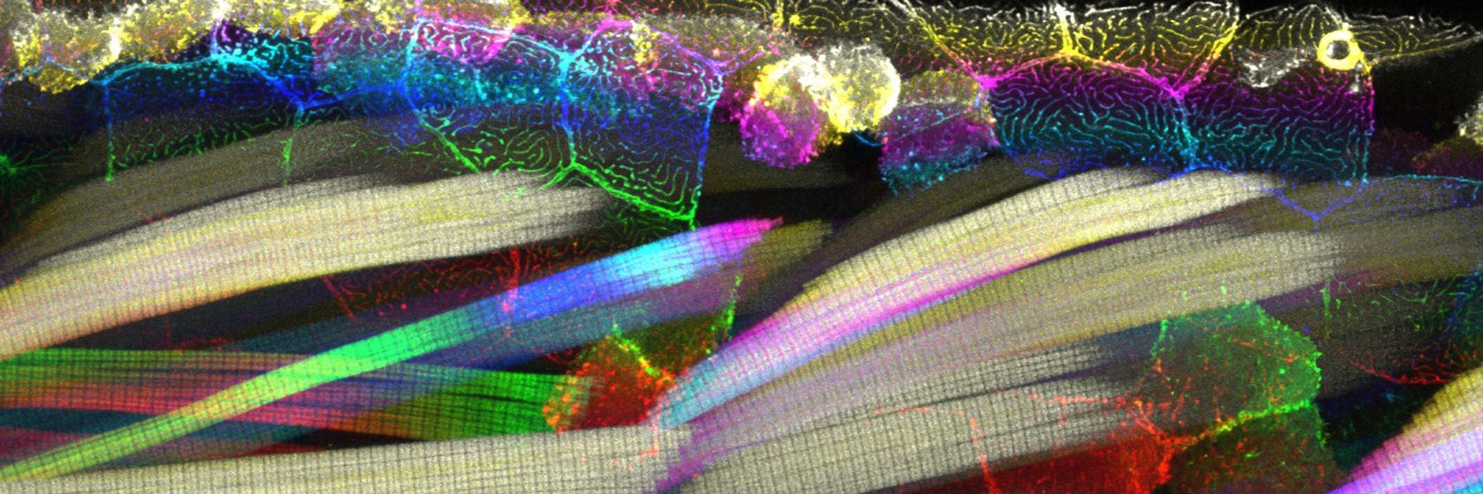
Is it just me or do these cells look a lot like the americas?
May 30, 2025 at 11:26 PM
Is it just me or do these cells look a lot like the americas?
FilaBuster - Vimentin IF fragmentation
May 25, 2025 at 3:47 AM
FilaBuster - Vimentin IF fragmentation
Happy to officially introduce FilaBuster - a strategy for rapid, light-mediated intermediate filament disassembly. Compatible with multiple IF types, modular in design, and precise enough to induce localized filament disassembly in live cells.
www.biorxiv.org/content/10.1...
www.biorxiv.org/content/10.1...
April 22, 2025 at 1:02 AM
Happy to officially introduce FilaBuster - a strategy for rapid, light-mediated intermediate filament disassembly. Compatible with multiple IF types, modular in design, and precise enough to induce localized filament disassembly in live cells.
www.biorxiv.org/content/10.1...
www.biorxiv.org/content/10.1...
In the paper, we introduce and characterize a new tool for tracking single vimentin IFs in live cells. After noticing single filaments moving independently within ostensibly tight bundles, we used FIB-SEM to take a closer look at vimentin bundle organization and vimentin-microtubule interactions.
March 10, 2025 at 6:49 PM
In the paper, we introduce and characterize a new tool for tracking single vimentin IFs in live cells. After noticing single filaments moving independently within ostensibly tight bundles, we used FIB-SEM to take a closer look at vimentin bundle organization and vimentin-microtubule interactions.
Poor little vimentin stick man 😱
A couple of months later than expected, but I'm finally gearing up to share *FilaBuster* - our approach for light-mediated IF disassembly. Stay tuned!
A couple of months later than expected, but I'm finally gearing up to share *FilaBuster* - our approach for light-mediated IF disassembly. Stay tuned!
March 10, 2025 at 12:53 AM
Poor little vimentin stick man 😱
A couple of months later than expected, but I'm finally gearing up to share *FilaBuster* - our approach for light-mediated IF disassembly. Stay tuned!
A couple of months later than expected, but I'm finally gearing up to share *FilaBuster* - our approach for light-mediated IF disassembly. Stay tuned!
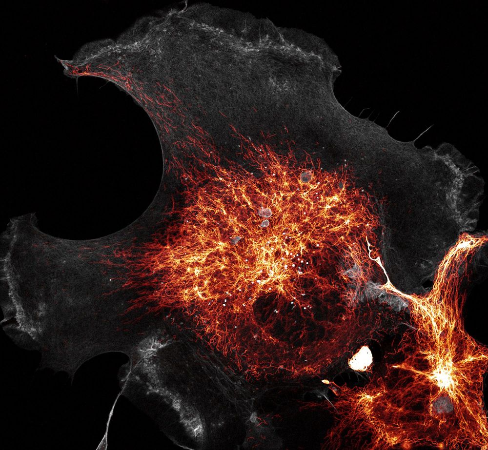
Vimentin (orange) and actin filaments (gray) in a COS-7. Airyscan images of the cytoskeleton always bring me joy. I square root transformed the vimentin so you can see the dimmer filaments out at the edge.
March 4, 2025 at 5:10 AM
Vimentin (orange) and actin filaments (gray) in a COS-7. Airyscan images of the cytoskeleton always bring me joy. I square root transformed the vimentin so you can see the dimmer filaments out at the edge.
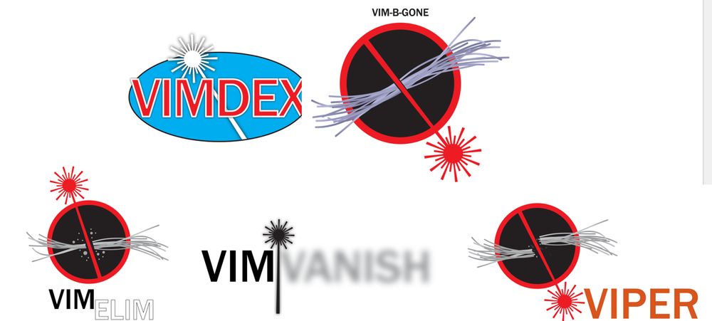
We're calling it FilaBuster (Filament Buster)
But I also spent (wasted?) a good amount of time designing fake logos and brainstorming alternative names.
But I also spent (wasted?) a good amount of time designing fake logos and brainstorming alternative names.

December 18, 2024 at 5:55 AM
We're calling it FilaBuster (Filament Buster)
But I also spent (wasted?) a good amount of time designing fake logos and brainstorming alternative names.
But I also spent (wasted?) a good amount of time designing fake logos and brainstorming alternative names.
Here's a still image of the effect (hopefully less compressed)

December 18, 2024 at 5:44 AM
Here's a still image of the effect (hopefully less compressed)
Really bummed to have missed ASCB this year due to some last-minute circumstances, but excited that our manuscript describing an IF disassembly tool will (hopefully) be posted soon. Here's a movie of vimentin color-coded by orientation.
December 18, 2024 at 5:40 AM
Really bummed to have missed ASCB this year due to some last-minute circumstances, but excited that our manuscript describing an IF disassembly tool will (hopefully) be posted soon. Here's a movie of vimentin color-coded by orientation.
The Cytoskeleton.... or at least some of it.
These are images of actin filaments (left), vimentin intermediate filaments (center), and microtubules (right) in COS7 cells.
These are images of actin filaments (left), vimentin intermediate filaments (center), and microtubules (right) in COS7 cells.

December 4, 2024 at 4:17 AM
The Cytoskeleton.... or at least some of it.
These are images of actin filaments (left), vimentin intermediate filaments (center), and microtubules (right) in COS7 cells.
These are images of actin filaments (left), vimentin intermediate filaments (center), and microtubules (right) in COS7 cells.
This is a movie of actin filaments in a cultured cell. I like watching it and I hope you do to.
November 18, 2024 at 5:50 AM
This is a movie of actin filaments in a cultured cell. I like watching it and I hope you do to.
1 second per frame for 6 minutes
Here is the colorcoded projection of the eb3 over 360 frames
Here is the colorcoded projection of the eb3 over 360 frames

November 15, 2024 at 6:10 AM
1 second per frame for 6 minutes
Here is the colorcoded projection of the eb3 over 360 frames
Here is the colorcoded projection of the eb3 over 360 frames
It's not a repost if it's a new platform, right? right??
Lattice light sheet movie of actin in mitosis. Blebby.
Lattice light sheet movie of actin in mitosis. Blebby.
November 15, 2024 at 6:04 AM
It's not a repost if it's a new platform, right? right??
Lattice light sheet movie of actin in mitosis. Blebby.
Lattice light sheet movie of actin in mitosis. Blebby.
FFmpeg spent substantially more time on this one. Let's see if the upload looks any better.
November 13, 2024 at 5:39 AM
FFmpeg spent substantially more time on this one. Let's see if the upload looks any better.
MP4 test. Please ignore.
These are intermediate filaments reconstructed from FIB-SEM but it doesn't matter. Let's say it's pasta. Just trying to work out which videos upload best.
These are intermediate filaments reconstructed from FIB-SEM but it doesn't matter. Let's say it's pasta. Just trying to work out which videos upload best.
November 13, 2024 at 5:38 AM
MP4 test. Please ignore.
These are intermediate filaments reconstructed from FIB-SEM but it doesn't matter. Let's say it's pasta. Just trying to work out which videos upload best.
These are intermediate filaments reconstructed from FIB-SEM but it doesn't matter. Let's say it's pasta. Just trying to work out which videos upload best.
OK swing and a miss on the GIF. Trying Mp4 again but smaller.
November 13, 2024 at 4:25 AM
OK swing and a miss on the GIF. Trying Mp4 again but smaller.