


Kevin Terretaz
@kwolbachia.bsky.social
Cells, microscopy, colorful LUTs and ImageJ
https://github.com/kwolbachia
For work I study Wolbachia symbiosis in Montpellier
https://github.com/kwolbachia
For work I study Wolbachia symbiosis in Montpellier
Pinned

My new ImageJ / Fiji toolkit is out 🔥! The goal is to make image handling & visualization easy, with an intuitive interface! Install it on Fiji with the "Image Viewer" update site
#microscopy #ImageJ #FluorescenceFriday #microscopyMonday
imagej.net/plugins/imag...
#microscopy #ImageJ #FluorescenceFriday #microscopyMonday
imagej.net/plugins/imag...
Reposted by Kevin Terretaz
*New preprint alert* uncovering a mechanical pacemaker that synchronizes nephron formation with branching of the kidney's epithelial tubule tree. Read below to learn about this twisty journey lead by Sam Grindel and Sachin Davis in the lab. [Movie by Nils Lindstrom]
www.biorxiv.org/content/10.1...
www.biorxiv.org/content/10.1...
December 13, 2025 at 12:08 AM
*New preprint alert* uncovering a mechanical pacemaker that synchronizes nephron formation with branching of the kidney's epithelial tubule tree. Read below to learn about this twisty journey lead by Sam Grindel and Sachin Davis in the lab. [Movie by Nils Lindstrom]
www.biorxiv.org/content/10.1...
www.biorxiv.org/content/10.1...
Reposted by Kevin Terretaz
This movie shows lysosomes (orange) and keratin (gray) in a cultured cell over 10 minutes.
December 12, 2025 at 6:20 AM
This movie shows lysosomes (orange) and keratin (gray) in a cultured cell over 10 minutes.
Reposted by Kevin Terretaz

A favorite recent image of some busy microglia, lovingly named the Rat King (yellow CD68, pink Iba1, blue DAPI) for my first #FluorescenceFriday

December 12, 2025 at 2:38 PM
A favorite recent image of some busy microglia, lovingly named the Rat King (yellow CD68, pink Iba1, blue DAPI) for my first #FluorescenceFriday
Reposted by Kevin Terretaz
A glittery, festive #FluorescenceFriday today, spent playing with some fun samples 🪩🕺
December 12, 2025 at 3:32 PM
A glittery, festive #FluorescenceFriday today, spent playing with some fun samples 🪩🕺
Reposted by Kevin Terretaz
Back in 2016, I spent some time centrifuging chambers containing a kinesin/microtubule -based active gel so that all the material ended up on one side of the chamber. Then recorded how long it took to expand.
December 3, 2025 at 2:49 PM
Back in 2016, I spent some time centrifuging chambers containing a kinesin/microtubule -based active gel so that all the material ended up on one side of the chamber. Then recorded how long it took to expand.
Reposted by Kevin Terretaz
Frog eye, frog eye. Can I have a confocal microscope to play on for 12 hrs a day again please? 👉👈🥺 🐸

December 9, 2025 at 7:58 PM
Frog eye, frog eye. Can I have a confocal microscope to play on for 12 hrs a day again please? 👉👈🥺 🐸
Unsolicited remix of some cool #FluorescenceFriday images from Matthias Böddener 😁can't go wrong with the Noice LUTS!
Really nice redesign of some of my pics by
@kwolbachia.bsky.social
thanks a lot for making them look great!
@kwolbachia.bsky.social
thanks a lot for making them look great!


December 9, 2025 at 4:17 PM
Unsolicited remix of some cool #FluorescenceFriday images from Matthias Böddener 😁can't go wrong with the Noice LUTS!
Reposted by Kevin Terretaz

The arbuscules of arbuscular mycorrhiza

December 9, 2025 at 5:57 AM
The arbuscules of arbuscular mycorrhiza
Reposted by Kevin Terretaz
#LivingArchitectures
We put cells and cytoskeleton filaments on the architecture of the musée d'Orsay.
www.musee-orsay.fr/fr/agenda/ev...
Scientists of the #CytoMorphoLab adapted their protocols to illustrate the questions that keep them awake at night.
-> Two shows on the 24th and 25th of January.
We put cells and cytoskeleton filaments on the architecture of the musée d'Orsay.
www.musee-orsay.fr/fr/agenda/ev...
Scientists of the #CytoMorphoLab adapted their protocols to illustrate the questions that keep them awake at night.
-> Two shows on the 24th and 25th of January.


December 5, 2025 at 4:11 PM
#LivingArchitectures
We put cells and cytoskeleton filaments on the architecture of the musée d'Orsay.
www.musee-orsay.fr/fr/agenda/ev...
Scientists of the #CytoMorphoLab adapted their protocols to illustrate the questions that keep them awake at night.
-> Two shows on the 24th and 25th of January.
We put cells and cytoskeleton filaments on the architecture of the musée d'Orsay.
www.musee-orsay.fr/fr/agenda/ev...
Scientists of the #CytoMorphoLab adapted their protocols to illustrate the questions that keep them awake at night.
-> Two shows on the 24th and 25th of January.
Reposted by Kevin Terretaz
Immunostaining of a mouse brain blood vessel with Collagen type 4 (cyan) and GFAP (orange) Nuclei have been visualized by DAPI staining (blue).
#FluorescenceFriday
#bloodbrainbarrier
#FluorescenceFriday
#bloodbrainbarrier
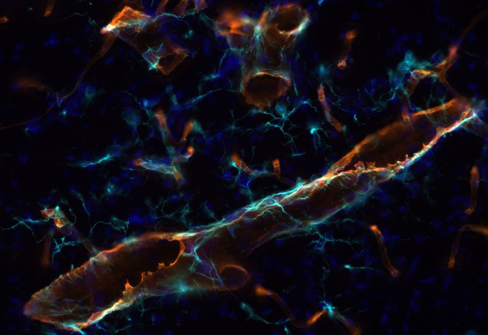
November 15, 2024 at 1:09 PM
Immunostaining of a mouse brain blood vessel with Collagen type 4 (cyan) and GFAP (orange) Nuclei have been visualized by DAPI staining (blue).
#FluorescenceFriday
#bloodbrainbarrier
#FluorescenceFriday
#bloodbrainbarrier
Reposted by Kevin Terretaz
This distal gill tip is giving full branchial art vibes for #FluorescenceFriday 🦓🐟
— who knew oxygen exchange could be this pretty.
🔴 kdrl:mCherry highlights the vasculature
⚪ fgf10b:nEOS marks the pillar cells
#Zebrafish #DevBio #SciArt
— who knew oxygen exchange could be this pretty.
🔴 kdrl:mCherry highlights the vasculature
⚪ fgf10b:nEOS marks the pillar cells
#Zebrafish #DevBio #SciArt

December 5, 2025 at 1:39 PM
This distal gill tip is giving full branchial art vibes for #FluorescenceFriday 🦓🐟
— who knew oxygen exchange could be this pretty.
🔴 kdrl:mCherry highlights the vasculature
⚪ fgf10b:nEOS marks the pillar cells
#Zebrafish #DevBio #SciArt
— who knew oxygen exchange could be this pretty.
🔴 kdrl:mCherry highlights the vasculature
⚪ fgf10b:nEOS marks the pillar cells
#Zebrafish #DevBio #SciArt
Reposted by Kevin Terretaz
New preprint! Do you like ocean waves? We found similar waves on bacterial colonies! We found that this collective behavior, known as rippling, is nothing but surface waves on an active nematic. @princeton.edu @mpipks.bsky.social @ub.edu @icreacommunity.bsky.social
www.biorxiv.org/content/10.1...
www.biorxiv.org/content/10.1...
December 2, 2025 at 8:02 PM
New preprint! Do you like ocean waves? We found similar waves on bacterial colonies! We found that this collective behavior, known as rippling, is nothing but surface waves on an active nematic. @princeton.edu @mpipks.bsky.social @ub.edu @icreacommunity.bsky.social
www.biorxiv.org/content/10.1...
www.biorxiv.org/content/10.1...
Reposted by Kevin Terretaz
Zebrafish tail depicting muscle and cell nuclei. Done at @cabd-upo-csic.bsky.social, in @gloriabreacalvo.bsky.social lab.
#FluorescenceFriday, #zebrafish, #DevelopmentalBiology,
#microscopy
#FluorescenceFriday, #zebrafish, #DevelopmentalBiology,
#microscopy

November 28, 2025 at 3:00 PM
Zebrafish tail depicting muscle and cell nuclei. Done at @cabd-upo-csic.bsky.social, in @gloriabreacalvo.bsky.social lab.
#FluorescenceFriday, #zebrafish, #DevelopmentalBiology,
#microscopy
#FluorescenceFriday, #zebrafish, #DevelopmentalBiology,
#microscopy
Reposted by Kevin Terretaz
Our M.Sc. PBioC (Physical Biology of Cells and Cell Interactions) students at @goetheuni.bsky.social had their first hands-on encounter with STED microscopy this week — and they captured some truly stunning super-resolution images.
#FluorescenceFriday #Superresolution
#FluorescenceFriday #Superresolution


November 28, 2025 at 11:59 PM
Our M.Sc. PBioC (Physical Biology of Cells and Cell Interactions) students at @goetheuni.bsky.social had their first hands-on encounter with STED microscopy this week — and they captured some truly stunning super-resolution images.
#FluorescenceFriday #Superresolution
#FluorescenceFriday #Superresolution
Reposted by Kevin Terretaz

For this week #FluorescenceFriday, we'll be treated with one of our favourite animal models 🐸 . Here is a Xenopus laevis embryo imaged with a Lightsheet microscope showing macrophage migration.
Purple🟣 = ectoderm nuclei, Red🔴 = macrophages.
📹: Hoang Anh Le, postdoc in the Mayor lab, UCL.
Purple🟣 = ectoderm nuclei, Red🔴 = macrophages.
📹: Hoang Anh Le, postdoc in the Mayor lab, UCL.
November 28, 2025 at 6:32 PM
For this week #FluorescenceFriday, we'll be treated with one of our favourite animal models 🐸 . Here is a Xenopus laevis embryo imaged with a Lightsheet microscope showing macrophage migration.
Purple🟣 = ectoderm nuclei, Red🔴 = macrophages.
📹: Hoang Anh Le, postdoc in the Mayor lab, UCL.
Purple🟣 = ectoderm nuclei, Red🔴 = macrophages.
📹: Hoang Anh Le, postdoc in the Mayor lab, UCL.
Reposted by Kevin Terretaz

I have more live imaging of actin in B cells from this week for this #FluorescenceFriday
November 28, 2025 at 1:39 PM
I have more live imaging of actin in B cells from this week for this #FluorescenceFriday
Reposted by Kevin Terretaz

So excited to share this as a new junior PI:
My brand-new lab website! 🎉🪰🌀
www.bischofflab.com
Please pass it on to young, motivated researchers looking for PhD positions 😊
And for the #FluorescenceFriday community: don’t miss the SciArt Gallery!
#CellBio #DevBio #PhDjob #PhDposition #Science
My brand-new lab website! 🎉🪰🌀
www.bischofflab.com
Please pass it on to young, motivated researchers looking for PhD positions 😊
And for the #FluorescenceFriday community: don’t miss the SciArt Gallery!
#CellBio #DevBio #PhDjob #PhDposition #Science


November 27, 2025 at 8:26 AM
So excited to share this as a new junior PI:
My brand-new lab website! 🎉🪰🌀
www.bischofflab.com
Please pass it on to young, motivated researchers looking for PhD positions 😊
And for the #FluorescenceFriday community: don’t miss the SciArt Gallery!
#CellBio #DevBio #PhDjob #PhDposition #Science
My brand-new lab website! 🎉🪰🌀
www.bischofflab.com
Please pass it on to young, motivated researchers looking for PhD positions 😊
And for the #FluorescenceFriday community: don’t miss the SciArt Gallery!
#CellBio #DevBio #PhDjob #PhDposition #Science
Reposted by Kevin Terretaz
Final result (better to do the rolling-ball background subtraction before merging, and merging using Max rather than blending)

November 26, 2025 at 4:30 PM
Final result (better to do the rolling-ball background subtraction before merging, and merging using Max rather than blending)
Reposted by Kevin Terretaz
So happy to announce our new preprint, “A geothermal amoeba sets a new upper temperature limit for eukaryotes.” We cultured a novel amoeba from Lassen Volcanic NP (CA, USA) that divides at 63°C (145°F) 🔥 - a new record for euk growth!
#protistsonsky 🧵
#protistsonsky 🧵
November 25, 2025 at 8:41 PM
So happy to announce our new preprint, “A geothermal amoeba sets a new upper temperature limit for eukaryotes.” We cultured a novel amoeba from Lassen Volcanic NP (CA, USA) that divides at 63°C (145°F) 🔥 - a new record for euk growth!
#protistsonsky 🧵
#protistsonsky 🧵
I finally got my hands on a MRI! Didn't help my wrist but now I have some really cool image stacks to play with 🤩
November 25, 2025 at 1:51 PM
I finally got my hands on a MRI! Didn't help my wrist but now I have some really cool image stacks to play with 🤩
Reposted by Kevin Terretaz
I like this movie, but a friend of mine likes to complain about the obvious stitching artifacts. I'll try harder next time, Michael. Vimentin (orange) and ER (blue) in an overnight acquisition.
November 25, 2025 at 6:06 AM
I like this movie, but a friend of mine likes to complain about the obvious stitching artifacts. I'll try harder next time, Michael. Vimentin (orange) and ER (blue) in an overnight acquisition.
Reposted by Kevin Terretaz
I gave a talk at Blender Conference yesterday, showing how biological volumes work, and how to visualize them in Blender 😋 youtu.be/WPajSWX730o?... #bcon25
Microscopy Nodes: handling large biological volumes — Blender Conference 2025
YouTube video by Blender
youtu.be
September 19, 2025 at 7:35 AM
I gave a talk at Blender Conference yesterday, showing how biological volumes work, and how to visualize them in Blender 😋 youtu.be/WPajSWX730o?... #bcon25
Reposted by Kevin Terretaz
Still posting cytoskeleton videos, it seems. Actin this time.
Sample: Lifeact-eGFP in HeLa cells.
Modality: Airyscan confocal
Timestamp is mm:ss and the scale bar is 5 µm.
Sample: Lifeact-eGFP in HeLa cells.
Modality: Airyscan confocal
Timestamp is mm:ss and the scale bar is 5 µm.
November 23, 2025 at 3:21 AM
Still posting cytoskeleton videos, it seems. Actin this time.
Sample: Lifeact-eGFP in HeLa cells.
Modality: Airyscan confocal
Timestamp is mm:ss and the scale bar is 5 µm.
Sample: Lifeact-eGFP in HeLa cells.
Modality: Airyscan confocal
Timestamp is mm:ss and the scale bar is 5 µm.
Reposted by Kevin Terretaz

iPS cell-derived cardiac myocytes (heart muscle cells) typically beat about once per second, so I usually speed up the movies I post; otherwise, scrollers might miss the action. But every now and then, a cell looks like this in real time. #CellBiology
November 10, 2025 at 1:56 AM
iPS cell-derived cardiac myocytes (heart muscle cells) typically beat about once per second, so I usually speed up the movies I post; otherwise, scrollers might miss the action. But every now and then, a cell looks like this in real time. #CellBiology
Reposted by Kevin Terretaz
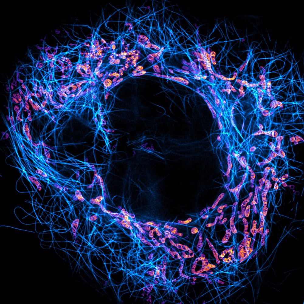
Attempt number seven at uploading this video of intermediate filaments in an enormous COS7 cell. I have a feeling the BlueSky compression will not do it any favors.
November 21, 2025 at 3:54 AM
Attempt number seven at uploading this video of intermediate filaments in an enormous COS7 cell. I have a feeling the BlueSky compression will not do it any favors.