


International Society of Developmental Biology
@isdb.bsky.social
ISDB is a non-profit scientific association that promotes the study of developmental biology
Pinned
Hello my lovely developmental biologists, The International Society for Developmental Biology is here with y'all in this #BlueSky. If you follow us on Twitter (we refuse to call it X), please drop us a follow here! Let's connect! #DevelopmentalBiology #cellbiology
Reposted by International Society of Developmental Biology
✨A gut feeling you’ll love this one 🐛 The anterior midgut of a Drosophila melanogaster 🪰larva puts on a confocal glow show! 🟢 Adult midgut progenitors 🔴 Actin 🔵 Nuclei 📸Image by Sapna Krishnakumar #FluorescenceFriday #Microscopy #DevBio

November 28, 2025 at 11:07 PM
✨A gut feeling you’ll love this one 🐛 The anterior midgut of a Drosophila melanogaster 🪰larva puts on a confocal glow show! 🟢 Adult midgut progenitors 🔴 Actin 🔵 Nuclei 📸Image by Sapna Krishnakumar #FluorescenceFriday #Microscopy #DevBio
Reposted by International Society of Developmental Biology
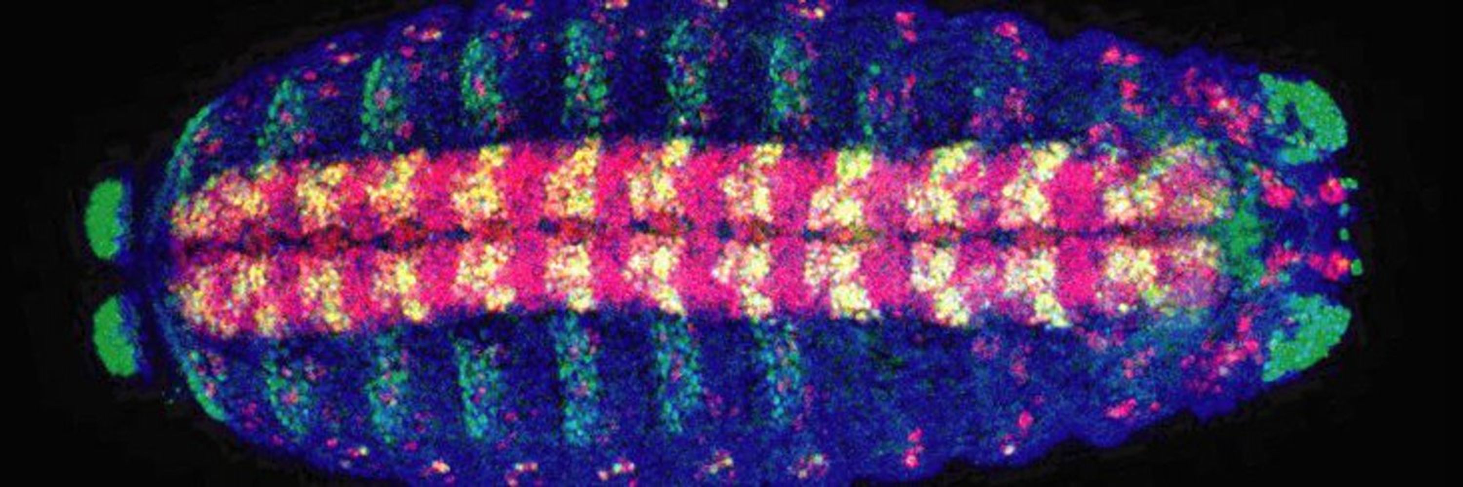
For this week #FluorescenceFriday, we'll be treated with one of our favourite animal models 🐸 . Here is a Xenopus laevis embryo imaged with a Lightsheet microscope showing macrophage migration.
Purple🟣 = ectoderm nuclei, Red🔴 = macrophages.
📹: Hoang Anh Le, postdoc in the Mayor lab, UCL.
Purple🟣 = ectoderm nuclei, Red🔴 = macrophages.
📹: Hoang Anh Le, postdoc in the Mayor lab, UCL.
November 28, 2025 at 6:32 PM
For this week #FluorescenceFriday, we'll be treated with one of our favourite animal models 🐸 . Here is a Xenopus laevis embryo imaged with a Lightsheet microscope showing macrophage migration.
Purple🟣 = ectoderm nuclei, Red🔴 = macrophages.
📹: Hoang Anh Le, postdoc in the Mayor lab, UCL.
Purple🟣 = ectoderm nuclei, Red🔴 = macrophages.
📹: Hoang Anh Le, postdoc in the Mayor lab, UCL.
Reposted by International Society of Developmental Biology
Stunning @anhhle2702.bsky.social 😍
For this week #FluorescenceFriday, we'll be treated with one of our favourite animal models 🐸 . Here is a Xenopus laevis embryo imaged with a Lightsheet microscope showing macrophage migration.
Purple🟣 = ectoderm nuclei, Red🔴 = macrophages.
📹: Hoang Anh Le, postdoc in the Mayor lab, UCL.
Purple🟣 = ectoderm nuclei, Red🔴 = macrophages.
📹: Hoang Anh Le, postdoc in the Mayor lab, UCL.
November 28, 2025 at 6:39 PM
Stunning @anhhle2702.bsky.social 😍
For this week #FluorescenceFriday, we'll be treated with one of our favourite animal models 🐸 . Here is a Xenopus laevis embryo imaged with a Lightsheet microscope showing macrophage migration.
Purple🟣 = ectoderm nuclei, Red🔴 = macrophages.
📹: Hoang Anh Le, postdoc in the Mayor lab, UCL.
Purple🟣 = ectoderm nuclei, Red🔴 = macrophages.
📹: Hoang Anh Le, postdoc in the Mayor lab, UCL.
November 28, 2025 at 6:32 PM
For this week #FluorescenceFriday, we'll be treated with one of our favourite animal models 🐸 . Here is a Xenopus laevis embryo imaged with a Lightsheet microscope showing macrophage migration.
Purple🟣 = ectoderm nuclei, Red🔴 = macrophages.
📹: Hoang Anh Le, postdoc in the Mayor lab, UCL.
Purple🟣 = ectoderm nuclei, Red🔴 = macrophages.
📹: Hoang Anh Le, postdoc in the Mayor lab, UCL.
Reposted by International Society of Developmental Biology

How do cytokines shape immune cells?
We analyzed and visualized Parse Bio's 10M PBMC dataset to give researchers a powerful tool for exploring the dynamics between cytokines & peripheral blood mononuclear cells.
More: https://apps.allenimmunology.org/aifi/resources/parse-10m-cytokines/
#ImmunoSky
We analyzed and visualized Parse Bio's 10M PBMC dataset to give researchers a powerful tool for exploring the dynamics between cytokines & peripheral blood mononuclear cells.
More: https://apps.allenimmunology.org/aifi/resources/parse-10m-cytokines/
#ImmunoSky

November 28, 2025 at 4:10 PM
How do cytokines shape immune cells?
We analyzed and visualized Parse Bio's 10M PBMC dataset to give researchers a powerful tool for exploring the dynamics between cytokines & peripheral blood mononuclear cells.
More: https://apps.allenimmunology.org/aifi/resources/parse-10m-cytokines/
#ImmunoSky
We analyzed and visualized Parse Bio's 10M PBMC dataset to give researchers a powerful tool for exploring the dynamics between cytokines & peripheral blood mononuclear cells.
More: https://apps.allenimmunology.org/aifi/resources/parse-10m-cytokines/
#ImmunoSky
Reposted by International Society of Developmental Biology

I have more live imaging of actin in B cells from this week for this #FluorescenceFriday
November 28, 2025 at 1:39 PM
I have more live imaging of actin in B cells from this week for this #FluorescenceFriday
Reposted by International Society of Developmental Biology
Registration deadline 20th Dec! We have generous travel grants for early career researchers! April is a beautiful time to visit China!
Our interdisciplinary @embo.org @biologists.bsky.social workshop on Biophysical and Molecular Mechanisms of Animal Homeostasis and Repair will take place in beautiful Haining, China, 6-10 April 2026. Exciting line up of international speakers! Register now: meetings.embo.org/event/26-hom...
Interfacing biophysical and molecular mechanisms of animal homeostasis and repair
The control of animal homeostasis and repair has been studied for decades, mostly from a molecular and biochemical perspective. Recent research shows that biophysical factors, such as mechanical forc…
meetings.embo.org
November 28, 2025 at 5:04 PM
Registration deadline 20th Dec! We have generous travel grants for early career researchers! April is a beautiful time to visit China!
Reposted by International Society of Developmental Biology
Microglial CLEC7A restrains amyloid beta plaque pathology in a mouse model of Alzheimer's disease
Fascinating study from the lab of @lukensjohnr.bsky.social
alz-journals.onlinelibrary.wiley.com/doi/10.1002/...
Fascinating study from the lab of @lukensjohnr.bsky.social
alz-journals.onlinelibrary.wiley.com/doi/10.1002/...

Microglial CLEC7A restrains amyloid beta plaque pathology in a mouse model of Alzheimer's disease
INTRODUCTION CLEC7A is a surface receptor that is highly upregulated on microglia in many Alzheimer's disease (AD) models. Little is known about the role that microglial CLEC7A signaling plays in AD...
alz-journals.onlinelibrary.wiley.com
November 28, 2025 at 6:06 PM
Microglial CLEC7A restrains amyloid beta plaque pathology in a mouse model of Alzheimer's disease
Fascinating study from the lab of @lukensjohnr.bsky.social
alz-journals.onlinelibrary.wiley.com/doi/10.1002/...
Fascinating study from the lab of @lukensjohnr.bsky.social
alz-journals.onlinelibrary.wiley.com/doi/10.1002/...
Reposted by International Society of Developmental Biology
Think about craniofacial biology in sunny California😎! Don't forget to submit an abstract for the 2026 GRC Craniofacial. Many talk slots available from abstract selections. Deadline for talk abstracts 14 Dec, coming soon.... ☠️🐭🐟🐸🧬 #craniofacial #genetics #neuralcrest www.grc.org/craniofacial...
2026 Craniofacial Morphogenesis and Tissue Regeneration Conference GRC
The 2026 Gordon Research Conference on Craniofacial Morphogenesis and Tissue Regeneration will be held in Ventura, California. Apply today to reserve your spot.
www.grc.org
November 28, 2025 at 5:39 PM
Think about craniofacial biology in sunny California😎! Don't forget to submit an abstract for the 2026 GRC Craniofacial. Many talk slots available from abstract selections. Deadline for talk abstracts 14 Dec, coming soon.... ☠️🐭🐟🐸🧬 #craniofacial #genetics #neuralcrest www.grc.org/craniofacial...
Reposted by International Society of Developmental Biology

Tissue-resident macrophages are doing more than just protecting from infection. In this interesting paper, René Fernando Abarca-Buis et al shows that they can promote reepithelialization and blastema formation and regulate the maturation of chondrocytes. Check it out here:
doi.org/10.1016/j.cd...
doi.org/10.1016/j.cd...


November 28, 2025 at 5:57 PM
Tissue-resident macrophages are doing more than just protecting from infection. In this interesting paper, René Fernando Abarca-Buis et al shows that they can promote reepithelialization and blastema formation and regulate the maturation of chondrocytes. Check it out here:
doi.org/10.1016/j.cd...
doi.org/10.1016/j.cd...
Reposted by International Society of Developmental Biology

Three more weeks to sign up!
#CellBio community, this one is for you! Applications are open for the #LakeConference on Modeling Life from Cells to Tissues – co-hosted with Circuit Neuroscience Basel!
📆 April 20-22, 2026
📍 Seattle, USA
🔬 All career stages welcomed.
Apply to attend by 12/12: alleninstitute.org/events/aics-...
📆 April 20-22, 2026
📍 Seattle, USA
🔬 All career stages welcomed.
Apply to attend by 12/12: alleninstitute.org/events/aics-...

2026 Lake Conference: Modeling Life from Cells to Tissues
Join the Allen Institute for Cell Science and Circuit Neuroscience Basel in Seattle, Washington, April 20–22, 2026, for a 3 day conference, Modeling...
alleninstitute.org
November 21, 2025 at 8:18 AM
Three more weeks to sign up!
Reposted by International Society of Developmental Biology
Happy #FluorescenceFriday. This beautiful video shows slice by slice section of a zebrafish embryo's head. The embryo is almost completely transparent, proving that zebrafish is an amazing model system for microscopy.
Brightfield, red = actin, blue = DAPI
📹: Postdoc Matyas BL (@Mongera lab, UCL)
Brightfield, red = actin, blue = DAPI
📹: Postdoc Matyas BL (@Mongera lab, UCL)
November 21, 2025 at 5:34 PM
Happy #FluorescenceFriday. This beautiful video shows slice by slice section of a zebrafish embryo's head. The embryo is almost completely transparent, proving that zebrafish is an amazing model system for microscopy.
Brightfield, red = actin, blue = DAPI
📹: Postdoc Matyas BL (@Mongera lab, UCL)
Brightfield, red = actin, blue = DAPI
📹: Postdoc Matyas BL (@Mongera lab, UCL)
Reposted by International Society of Developmental Biology
We love zebrafish too! <3 #FluorescenceFriday #devbio
Happy #FluorescenceFriday. This beautiful video shows slice by slice section of a zebrafish embryo's head. The embryo is almost completely transparent, proving that zebrafish is an amazing model system for microscopy.
Brightfield, red = actin, blue = DAPI
📹: Postdoc Matyas BL (@Mongera lab, UCL)
Brightfield, red = actin, blue = DAPI
📹: Postdoc Matyas BL (@Mongera lab, UCL)
November 21, 2025 at 5:36 PM
We love zebrafish too! <3 #FluorescenceFriday #devbio
Reposted by International Society of Developmental Biology
It is not often that Sialoglycoproteins and sialyltransferases are mentioned during early development. This interesting review from the lab of Katia Cailliau brings the interesting biology of sialic acid into the context of #devbio, particularly during blastula formation.
doi.org/10.1016/j.cd...
doi.org/10.1016/j.cd...


November 21, 2025 at 5:46 PM
It is not often that Sialoglycoproteins and sialyltransferases are mentioned during early development. This interesting review from the lab of Katia Cailliau brings the interesting biology of sialic acid into the context of #devbio, particularly during blastula formation.
doi.org/10.1016/j.cd...
doi.org/10.1016/j.cd...
Happy #FluorescenceFriday. This beautiful video shows slice by slice section of a zebrafish embryo's head. The embryo is almost completely transparent, proving that zebrafish is an amazing model system for microscopy.
Brightfield, red = actin, blue = DAPI
📹: Postdoc Matyas BL (@Mongera lab, UCL)
Brightfield, red = actin, blue = DAPI
📹: Postdoc Matyas BL (@Mongera lab, UCL)
November 21, 2025 at 5:34 PM
Happy #FluorescenceFriday. This beautiful video shows slice by slice section of a zebrafish embryo's head. The embryo is almost completely transparent, proving that zebrafish is an amazing model system for microscopy.
Brightfield, red = actin, blue = DAPI
📹: Postdoc Matyas BL (@Mongera lab, UCL)
Brightfield, red = actin, blue = DAPI
📹: Postdoc Matyas BL (@Mongera lab, UCL)
Reposted by International Society of Developmental Biology

☕The authors identify a chemical cocktail to generate #totipotent - like cells, which they then use to build an #embryo model. This model captures a developmental spectrum from early #embryogenesis to post-implantation events.
bit.ly/4oHxUZp
bit.ly/4oHxUZp

A continuous totipotent-like cell-based embryo model recapitulates mouse embryogenesis from zygotic genome activation to gastrulation - Nature Cell Biology
The authors identify a chemical cocktail to generate totipotent-like cells, which they then use to build an embryo model. This model captures a developmental spectrum from early embryogenesis to post-...
bit.ly
November 15, 2025 at 8:17 PM
☕The authors identify a chemical cocktail to generate #totipotent - like cells, which they then use to build an #embryo model. This model captures a developmental spectrum from early #embryogenesis to post-implantation events.
bit.ly/4oHxUZp
bit.ly/4oHxUZp
Reposted by International Society of Developmental Biology
This week's #FluorescenceFriday, we'll be treated with one of the classical models of #devbio, the neural crest cells. Here is a beautiful video of neural crest cells with GFP-tagged focal adhesion kinase (🔵) and LifeAct-RFP (🟣) migrating on a Fibronectin matrix.
📹: Adam Shellard
📹: Adam Shellard
November 14, 2025 at 5:45 PM
This week's #FluorescenceFriday, we'll be treated with one of the classical models of #devbio, the neural crest cells. Here is a beautiful video of neural crest cells with GFP-tagged focal adhesion kinase (🔵) and LifeAct-RFP (🟣) migrating on a Fibronectin matrix.
📹: Adam Shellard
📹: Adam Shellard
This week's #FluorescenceFriday, we'll be treated with one of the classical models of #devbio, the neural crest cells. Here is a beautiful video of neural crest cells with GFP-tagged focal adhesion kinase (🔵) and LifeAct-RFP (🟣) migrating on a Fibronectin matrix.
📹: Adam Shellard
📹: Adam Shellard
November 14, 2025 at 5:45 PM
This week's #FluorescenceFriday, we'll be treated with one of the classical models of #devbio, the neural crest cells. Here is a beautiful video of neural crest cells with GFP-tagged focal adhesion kinase (🔵) and LifeAct-RFP (🟣) migrating on a Fibronectin matrix.
📹: Adam Shellard
📹: Adam Shellard
Reposted by International Society of Developmental Biology
Call for papers: Chilean Developmental Biology: at Home and Around the World - Cells & Development.
If you're a Chile-based scientist and Chilean researcher working abroad, please consider submit to us for this special issue.
‼️Submission deadline: 30 June 2026
www.sciencedirect.com/special-issu...
If you're a Chile-based scientist and Chilean researcher working abroad, please consider submit to us for this special issue.
‼️Submission deadline: 30 June 2026
www.sciencedirect.com/special-issu...
November 14, 2025 at 5:31 PM
Call for papers: Chilean Developmental Biology: at Home and Around the World - Cells & Development.
If you're a Chile-based scientist and Chilean researcher working abroad, please consider submit to us for this special issue.
‼️Submission deadline: 30 June 2026
www.sciencedirect.com/special-issu...
If you're a Chile-based scientist and Chilean researcher working abroad, please consider submit to us for this special issue.
‼️Submission deadline: 30 June 2026
www.sciencedirect.com/special-issu...
Reposted by International Society of Developmental Biology

We are pleased to announce our next #SEBD2026 meeting! The meeting will take place in El Rompido, Huelva, from the 28th to the 30th of October 2026 #savethedate @cabd-upo-csic.bsky.social @isdb.bsky.social @gfeev.bsky.social @ijdb.bsky.social @devbiol.bsky.social @devdynamics.bsky.social

November 14, 2025 at 7:53 AM
We are pleased to announce our next #SEBD2026 meeting! The meeting will take place in El Rompido, Huelva, from the 28th to the 30th of October 2026 #savethedate @cabd-upo-csic.bsky.social @isdb.bsky.social @gfeev.bsky.social @ijdb.bsky.social @devbiol.bsky.social @devdynamics.bsky.social
Reposted by International Society of Developmental Biology
Meis2 is a transcriptional factor involved in neurodevelopment. This paper characterises its distribution in the developing brain of Xenopus leavis. Check it out!
🚨✨🐸 Very excited to share the latest work I’m part of:
“Developmental and adult expression of the Meis2 transcription factor in the CNS of Xenopus laevis”.
Read below 👇
DOI: doi.org/10.3389/fnan...
#Neurogenesis #BrainDevelopment #Neuroscience #EvoDevo #DevBio #Xenopus
“Developmental and adult expression of the Meis2 transcription factor in the CNS of Xenopus laevis”.
Read below 👇
DOI: doi.org/10.3389/fnan...
#Neurogenesis #BrainDevelopment #Neuroscience #EvoDevo #DevBio #Xenopus


November 12, 2025 at 12:15 PM
Meis2 is a transcriptional factor involved in neurodevelopment. This paper characterises its distribution in the developing brain of Xenopus leavis. Check it out!
Meis2 is a transcriptional factor involved in neurodevelopment. This paper characterises its distribution in the developing brain of Xenopus leavis. Check it out!
🚨✨🐸 Very excited to share the latest work I’m part of:
“Developmental and adult expression of the Meis2 transcription factor in the CNS of Xenopus laevis”.
Read below 👇
DOI: doi.org/10.3389/fnan...
#Neurogenesis #BrainDevelopment #Neuroscience #EvoDevo #DevBio #Xenopus
“Developmental and adult expression of the Meis2 transcription factor in the CNS of Xenopus laevis”.
Read below 👇
DOI: doi.org/10.3389/fnan...
#Neurogenesis #BrainDevelopment #Neuroscience #EvoDevo #DevBio #Xenopus
November 12, 2025 at 12:15 PM
Meis2 is a transcriptional factor involved in neurodevelopment. This paper characterises its distribution in the developing brain of Xenopus leavis. Check it out!
Reposted by International Society of Developmental Biology

🚨✨🐸 Very excited to share the latest work I’m part of:
“Developmental and adult expression of the Meis2 transcription factor in the CNS of Xenopus laevis”.
Read below 👇
DOI: doi.org/10.3389/fnan...
#Neurogenesis #BrainDevelopment #Neuroscience #EvoDevo #DevBio #Xenopus
“Developmental and adult expression of the Meis2 transcription factor in the CNS of Xenopus laevis”.
Read below 👇
DOI: doi.org/10.3389/fnan...
#Neurogenesis #BrainDevelopment #Neuroscience #EvoDevo #DevBio #Xenopus
November 10, 2025 at 9:35 AM
🚨✨🐸 Very excited to share the latest work I’m part of:
“Developmental and adult expression of the Meis2 transcription factor in the CNS of Xenopus laevis”.
Read below 👇
DOI: doi.org/10.3389/fnan...
#Neurogenesis #BrainDevelopment #Neuroscience #EvoDevo #DevBio #Xenopus
“Developmental and adult expression of the Meis2 transcription factor in the CNS of Xenopus laevis”.
Read below 👇
DOI: doi.org/10.3389/fnan...
#Neurogenesis #BrainDevelopment #Neuroscience #EvoDevo #DevBio #Xenopus
Reposted by International Society of Developmental Biology
Sir John Gurdon was also the president of the @isdb.bsky.social from 1989-1992. He also facilitated or influenced the establishment of the @bsdb.bsky.social, @lasdb.bsky.social, many institutes, and generations of scientists, and worked with @biologists.bsky.social for decades. You will be missed.
"His secret weapons were an unquenchable curiosity and continuing hands-on experimentation at the bench with his beautiful frog embryos and oocytes".
A thoughtful tribute to the life of Sir John Gurdon by Edward M. De Robertis that we really recommend everyone to read.
doi.org/10.1016/j.cd...
A thoughtful tribute to the life of Sir John Gurdon by Edward M. De Robertis that we really recommend everyone to read.
doi.org/10.1016/j.cd...

November 7, 2025 at 9:23 PM
Sir John Gurdon was also the president of the @isdb.bsky.social from 1989-1992. He also facilitated or influenced the establishment of the @bsdb.bsky.social, @lasdb.bsky.social, many institutes, and generations of scientists, and worked with @biologists.bsky.social for decades. You will be missed.
Sir John Gurdon was also the president of the @isdb.bsky.social from 1989-1992. He also facilitated or influenced the establishment of the @bsdb.bsky.social, @lasdb.bsky.social, many institutes, and generations of scientists, and worked with @biologists.bsky.social for decades. You will be missed.
"His secret weapons were an unquenchable curiosity and continuing hands-on experimentation at the bench with his beautiful frog embryos and oocytes".
A thoughtful tribute to the life of Sir John Gurdon by Edward M. De Robertis that we really recommend everyone to read.
doi.org/10.1016/j.cd...
A thoughtful tribute to the life of Sir John Gurdon by Edward M. De Robertis that we really recommend everyone to read.
doi.org/10.1016/j.cd...
November 7, 2025 at 9:23 PM
Sir John Gurdon was also the president of the @isdb.bsky.social from 1989-1992. He also facilitated or influenced the establishment of the @bsdb.bsky.social, @lasdb.bsky.social, many institutes, and generations of scientists, and worked with @biologists.bsky.social for decades. You will be missed.