


Mathieu Preußner
@mathpreu.bsky.social
PhD in the lab of @vlecaudey.bsky.social at JGU. Developmental Biology in #Zebrafish.
The pronephric ducts 😉

November 14, 2025 at 1:33 PM
The pronephric ducts 😉
#FluorescenceFriday – Kidney Edition!
Giving the gills a day off 😉
Cryosection of 5dpf old 🦓🐟 larvae showing:
🟡podocytes
🟢VitaminD-binding protein-GFP
⚪Phalloidin
Notice how there’s no GFP signal in the pronephric duct — proof that filtration barrier is working perfectly
#Zebrafish #DevBio
Giving the gills a day off 😉
Cryosection of 5dpf old 🦓🐟 larvae showing:
🟡podocytes
🟢VitaminD-binding protein-GFP
⚪Phalloidin
Notice how there’s no GFP signal in the pronephric duct — proof that filtration barrier is working perfectly
#Zebrafish #DevBio

November 14, 2025 at 1:33 PM
#FluorescenceFriday – Kidney Edition!
Giving the gills a day off 😉
Cryosection of 5dpf old 🦓🐟 larvae showing:
🟡podocytes
🟢VitaminD-binding protein-GFP
⚪Phalloidin
Notice how there’s no GFP signal in the pronephric duct — proof that filtration barrier is working perfectly
#Zebrafish #DevBio
Giving the gills a day off 😉
Cryosection of 5dpf old 🦓🐟 larvae showing:
🟡podocytes
🟢VitaminD-binding protein-GFP
⚪Phalloidin
Notice how there’s no GFP signal in the pronephric duct — proof that filtration barrier is working perfectly
#Zebrafish #DevBio
Can't believe it — my first‑author paper is out and my image graces the cover of @dev-journal.bsky.social 🎉
Here, we reveal how early developmental programs shape and maintain #zebrafish gill architecture throughout life
🔗 journals.biologists.com/dev/issue/15...
#FluorescenceFriday #LifelongDevSI
Here, we reveal how early developmental programs shape and maintain #zebrafish gill architecture throughout life
🔗 journals.biologists.com/dev/issue/15...
#FluorescenceFriday #LifelongDevSI

October 31, 2025 at 10:55 AM
Can't believe it — my first‑author paper is out and my image graces the cover of @dev-journal.bsky.social 🎉
Here, we reveal how early developmental programs shape and maintain #zebrafish gill architecture throughout life
🔗 journals.biologists.com/dev/issue/15...
#FluorescenceFriday #LifelongDevSI
Here, we reveal how early developmental programs shape and maintain #zebrafish gill architecture throughout life
🔗 journals.biologists.com/dev/issue/15...
#FluorescenceFriday #LifelongDevSI
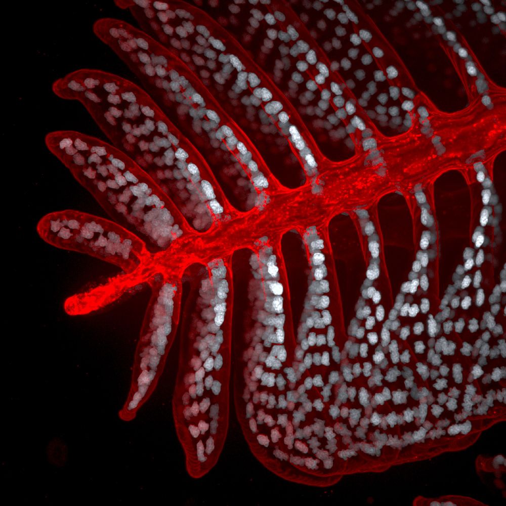
For this #FluorescenceFriday 🔬
a cryosection of a dissected zebrafish gill, stained with phalloidin (orange) and DAPI (blue-ish).
#Zebrafish #DevBio #Microscopy
a cryosection of a dissected zebrafish gill, stained with phalloidin (orange) and DAPI (blue-ish).
#Zebrafish #DevBio #Microscopy

October 17, 2025 at 3:15 PM
For this #FluorescenceFriday 🔬
a cryosection of a dissected zebrafish gill, stained with phalloidin (orange) and DAPI (blue-ish).
#Zebrafish #DevBio #Microscopy
a cryosection of a dissected zebrafish gill, stained with phalloidin (orange) and DAPI (blue-ish).
#Zebrafish #DevBio #Microscopy
15🧵
For those who made it this far... 👀
We’re not done with pillar cells.
They’ve got a lot more to say — about mechanics, structure, and dynamics.
More soon in our next paper. Stay tuned! 🧠🧪🔬
AGAIN! Big thanks to everyone involved—especially to
@vlecaudey.bsky.social
For those who made it this far... 👀
We’re not done with pillar cells.
They’ve got a lot more to say — about mechanics, structure, and dynamics.
More soon in our next paper. Stay tuned! 🧠🧪🔬
AGAIN! Big thanks to everyone involved—especially to
@vlecaudey.bsky.social

September 17, 2025 at 1:59 PM
15🧵
For those who made it this far... 👀
We’re not done with pillar cells.
They’ve got a lot more to say — about mechanics, structure, and dynamics.
More soon in our next paper. Stay tuned! 🧠🧪🔬
AGAIN! Big thanks to everyone involved—especially to
@vlecaudey.bsky.social
For those who made it this far... 👀
We’re not done with pillar cells.
They’ve got a lot more to say — about mechanics, structure, and dynamics.
More soon in our next paper. Stay tuned! 🧠🧪🔬
AGAIN! Big thanks to everyone involved—especially to
@vlecaudey.bsky.social
14🧵
Gills offer a powerful model to study :
🩸 Angiogenesis
💨 Blood flow & pressure
🧠 Neural crest stem cells
🌿 Branching morphogenesis — all in vivo.
Our work provides a detailed framework to explore these complex processes.
Gills offer a powerful model to study :
🩸 Angiogenesis
💨 Blood flow & pressure
🧠 Neural crest stem cells
🌿 Branching morphogenesis — all in vivo.
Our work provides a detailed framework to explore these complex processes.
September 17, 2025 at 1:59 PM
14🧵
Gills offer a powerful model to study :
🩸 Angiogenesis
💨 Blood flow & pressure
🧠 Neural crest stem cells
🌿 Branching morphogenesis — all in vivo.
Our work provides a detailed framework to explore these complex processes.
Gills offer a powerful model to study :
🩸 Angiogenesis
💨 Blood flow & pressure
🧠 Neural crest stem cells
🌿 Branching morphogenesis — all in vivo.
Our work provides a detailed framework to explore these complex processes.
13🧵
We quantified pillar cell numbers using:
🔬 ECI-based tissue clearing
📏 Isolation of single lamellae from filaments
Then, with the IMARIS Spots Module, we extracted detailed spatial and quantitative data — giving us a precise map of pillar cell distribution.
We quantified pillar cell numbers using:
🔬 ECI-based tissue clearing
📏 Isolation of single lamellae from filaments
Then, with the IMARIS Spots Module, we extracted detailed spatial and quantitative data — giving us a precise map of pillar cell distribution.
September 17, 2025 at 1:59 PM
13🧵
We quantified pillar cell numbers using:
🔬 ECI-based tissue clearing
📏 Isolation of single lamellae from filaments
Then, with the IMARIS Spots Module, we extracted detailed spatial and quantitative data — giving us a precise map of pillar cell distribution.
We quantified pillar cell numbers using:
🔬 ECI-based tissue clearing
📏 Isolation of single lamellae from filaments
Then, with the IMARIS Spots Module, we extracted detailed spatial and quantitative data — giving us a precise map of pillar cell distribution.
12🧵
Pillar cell number and lamella size showed clear developmental asymmetries — these also depend on:
📍 Whether the filament is medial or lateral
📍 Its position along the dorsoventral axis
This structural asymmetry likely reflects different functional roles of filaments and lamellae.
Pillar cell number and lamella size showed clear developmental asymmetries — these also depend on:
📍 Whether the filament is medial or lateral
📍 Its position along the dorsoventral axis
This structural asymmetry likely reflects different functional roles of filaments and lamellae.

September 17, 2025 at 1:59 PM
12🧵
Pillar cell number and lamella size showed clear developmental asymmetries — these also depend on:
📍 Whether the filament is medial or lateral
📍 Its position along the dorsoventral axis
This structural asymmetry likely reflects different functional roles of filaments and lamellae.
Pillar cell number and lamella size showed clear developmental asymmetries — these also depend on:
📍 Whether the filament is medial or lateral
📍 Its position along the dorsoventral axis
This structural asymmetry likely reflects different functional roles of filaments and lamellae.
11🧵
But endothelial cells don’t work alone.
Lamellae morphogenesis depends on tight coordination between:
🔴Endothelial cells
⚪Pillar cells (neural crest-derived!)
But endothelial cells don’t work alone.
Lamellae morphogenesis depends on tight coordination between:
🔴Endothelial cells
⚪Pillar cells (neural crest-derived!)

September 17, 2025 at 1:59 PM
11🧵
But endothelial cells don’t work alone.
Lamellae morphogenesis depends on tight coordination between:
🔴Endothelial cells
⚪Pillar cells (neural crest-derived!)
But endothelial cells don’t work alone.
Lamellae morphogenesis depends on tight coordination between:
🔴Endothelial cells
⚪Pillar cells (neural crest-derived!)
10🧵
Now, lets talk more about lamellae 🍂
We examined the development of gill lamellae.
It all started with an endothelial tip cell from the efferent filamental artery, which migrated across the lamella toward the low oxygen afferent side.
➡️ This forms the outer marginal channel (OMC)
Now, lets talk more about lamellae 🍂
We examined the development of gill lamellae.
It all started with an endothelial tip cell from the efferent filamental artery, which migrated across the lamella toward the low oxygen afferent side.
➡️ This forms the outer marginal channel (OMC)

September 17, 2025 at 1:59 PM
10🧵
Now, lets talk more about lamellae 🍂
We examined the development of gill lamellae.
It all started with an endothelial tip cell from the efferent filamental artery, which migrated across the lamella toward the low oxygen afferent side.
➡️ This forms the outer marginal channel (OMC)
Now, lets talk more about lamellae 🍂
We examined the development of gill lamellae.
It all started with an endothelial tip cell from the efferent filamental artery, which migrated across the lamella toward the low oxygen afferent side.
➡️ This forms the outer marginal channel (OMC)
Medial filaments were generally longer with more lamellae along most of the dorsoventral axis — except ventrally, where lateral filaments took the lead.
The asymmetry reflects developmental timing: medial filaments form first dorsally, lateral ones ventrally — shaping function well into adulthood.
The asymmetry reflects developmental timing: medial filaments form first dorsally, lateral ones ventrally — shaping function well into adulthood.

September 17, 2025 at 1:59 PM
Medial filaments were generally longer with more lamellae along most of the dorsoventral axis — except ventrally, where lateral filaments took the lead.
The asymmetry reflects developmental timing: medial filaments form first dorsally, lateral ones ventrally — shaping function well into adulthood.
The asymmetry reflects developmental timing: medial filaments form first dorsally, lateral ones ventrally — shaping function well into adulthood.
9🧵
This specific early gill developmental pattern sets the stage for adult gill architecture.
We tracked filament and lamella number and distribution across the 4 gill arches between 3.5 and 14.5 dpf up to 6 month.
This specific early gill developmental pattern sets the stage for adult gill architecture.
We tracked filament and lamella number and distribution across the 4 gill arches between 3.5 and 14.5 dpf up to 6 month.

September 17, 2025 at 1:59 PM
9🧵
This specific early gill developmental pattern sets the stage for adult gill architecture.
We tracked filament and lamella number and distribution across the 4 gill arches between 3.5 and 14.5 dpf up to 6 month.
This specific early gill developmental pattern sets the stage for adult gill architecture.
We tracked filament and lamella number and distribution across the 4 gill arches between 3.5 and 14.5 dpf up to 6 month.
8🧵
But then something unexpected happened (4–5 dpf).
Branching of the medial branchial artery marked a key symmetry-breaking event that separated how filaments formed dorsally vs. ventrally.
👇 In the ventral region (below MBA branch):
🔹 Lateral filaments sprout first
🔹 Medial filaments form later
But then something unexpected happened (4–5 dpf).
Branching of the medial branchial artery marked a key symmetry-breaking event that separated how filaments formed dorsally vs. ventrally.
👇 In the ventral region (below MBA branch):
🔹 Lateral filaments sprout first
🔹 Medial filaments form later

September 17, 2025 at 1:59 PM
8🧵
But then something unexpected happened (4–5 dpf).
Branching of the medial branchial artery marked a key symmetry-breaking event that separated how filaments formed dorsally vs. ventrally.
👇 In the ventral region (below MBA branch):
🔹 Lateral filaments sprout first
🔹 Medial filaments form later
But then something unexpected happened (4–5 dpf).
Branching of the medial branchial artery marked a key symmetry-breaking event that separated how filaments formed dorsally vs. ventrally.
👇 In the ventral region (below MBA branch):
🔹 Lateral filaments sprout first
🔹 Medial filaments form later
⚪lateral filament formation
September 17, 2025 at 1:59 PM
⚪lateral filament formation
⚪medial filament formation
September 17, 2025 at 1:59 PM
⚪medial filament formation
7🧵
In the dorsal area of the arch, medial filaments emerged first and extended the LBA by connecting to their neighbouring medial filaments. Only after this connection was made, lateral filaments emerged and fused with the LBA.
➡️ We used live imaging to show this formation in action.
In the dorsal area of the arch, medial filaments emerged first and extended the LBA by connecting to their neighbouring medial filaments. Only after this connection was made, lateral filaments emerged and fused with the LBA.
➡️ We used live imaging to show this formation in action.

September 17, 2025 at 1:59 PM
7🧵
In the dorsal area of the arch, medial filaments emerged first and extended the LBA by connecting to their neighbouring medial filaments. Only after this connection was made, lateral filaments emerged and fused with the LBA.
➡️ We used live imaging to show this formation in action.
In the dorsal area of the arch, medial filaments emerged first and extended the LBA by connecting to their neighbouring medial filaments. Only after this connection was made, lateral filaments emerged and fused with the LBA.
➡️ We used live imaging to show this formation in action.
6🧵
Once the newly established lateral branchial artery (LBA) fused with the branchial artery (BA), the segment of the original BA, located above the fusion site, will be designated as the medial branchial artery (MBA).
Once the newly established lateral branchial artery (LBA) fused with the branchial artery (BA), the segment of the original BA, located above the fusion site, will be designated as the medial branchial artery (MBA).

September 17, 2025 at 1:59 PM
6🧵
Once the newly established lateral branchial artery (LBA) fused with the branchial artery (BA), the segment of the original BA, located above the fusion site, will be designated as the medial branchial artery (MBA).
Once the newly established lateral branchial artery (LBA) fused with the branchial artery (BA), the segment of the original BA, located above the fusion site, will be designated as the medial branchial artery (MBA).
5🧵
So when do gill filaments form?
Using high resolution live-cell spinning disc microscopy, we mapped their onset.
The first medial filaments appeared around 2.5 dpf. They sprouted from the branchial artery (BA) and began connecting to neighbours — forming the lateral branchial artery (LBA).
So when do gill filaments form?
Using high resolution live-cell spinning disc microscopy, we mapped their onset.
The first medial filaments appeared around 2.5 dpf. They sprouted from the branchial artery (BA) and began connecting to neighbours — forming the lateral branchial artery (LBA).

September 17, 2025 at 1:59 PM
5🧵
So when do gill filaments form?
Using high resolution live-cell spinning disc microscopy, we mapped their onset.
The first medial filaments appeared around 2.5 dpf. They sprouted from the branchial artery (BA) and began connecting to neighbours — forming the lateral branchial artery (LBA).
So when do gill filaments form?
Using high resolution live-cell spinning disc microscopy, we mapped their onset.
The first medial filaments appeared around 2.5 dpf. They sprouted from the branchial artery (BA) and began connecting to neighbours — forming the lateral branchial artery (LBA).
4🧵
Let’s zoom in 🔍
To study filament morphology in more detail, we used two transgenic markers:
🔴 kdrl:mCherry — marks endothelial vasculature
⚪ fgf10b:nEOS — highlights pillar cell nuclei within the lamellae
Big thanks to @fabianlab.bsky.social for the fgf10b:nEOS line
Let’s zoom in 🔍
To study filament morphology in more detail, we used two transgenic markers:
🔴 kdrl:mCherry — marks endothelial vasculature
⚪ fgf10b:nEOS — highlights pillar cell nuclei within the lamellae
Big thanks to @fabianlab.bsky.social for the fgf10b:nEOS line

September 17, 2025 at 1:59 PM
4🧵
Let’s zoom in 🔍
To study filament morphology in more detail, we used two transgenic markers:
🔴 kdrl:mCherry — marks endothelial vasculature
⚪ fgf10b:nEOS — highlights pillar cell nuclei within the lamellae
Big thanks to @fabianlab.bsky.social for the fgf10b:nEOS line
Let’s zoom in 🔍
To study filament morphology in more detail, we used two transgenic markers:
🔴 kdrl:mCherry — marks endothelial vasculature
⚪ fgf10b:nEOS — highlights pillar cell nuclei within the lamellae
Big thanks to @fabianlab.bsky.social for the fgf10b:nEOS line
3🧵
Quick primer: Zebrafish have 4 gill arches on each side with filaments. Each filament harbours several stacked lamellae: the site of gas exchange.
Each lamella is encircled by the endothelial outer marginal channel (OMC) and supported by many pillar cells, which maintain the intergrity.
Quick primer: Zebrafish have 4 gill arches on each side with filaments. Each filament harbours several stacked lamellae: the site of gas exchange.
Each lamella is encircled by the endothelial outer marginal channel (OMC) and supported by many pillar cells, which maintain the intergrity.

September 17, 2025 at 1:59 PM
3🧵
Quick primer: Zebrafish have 4 gill arches on each side with filaments. Each filament harbours several stacked lamellae: the site of gas exchange.
Each lamella is encircled by the endothelial outer marginal channel (OMC) and supported by many pillar cells, which maintain the intergrity.
Quick primer: Zebrafish have 4 gill arches on each side with filaments. Each filament harbours several stacked lamellae: the site of gas exchange.
Each lamella is encircled by the endothelial outer marginal channel (OMC) and supported by many pillar cells, which maintain the intergrity.
2🧵
This curiosity-driven project started by sheer coincidence. While imaging the gills for a different scientific question, I noticed a consistent pattern: filament length varied along the dorsoventral axis.
🔴 Medial filaments (red) were longer than
⚪ Lateral filaments (white) —except ventrally.
This curiosity-driven project started by sheer coincidence. While imaging the gills for a different scientific question, I noticed a consistent pattern: filament length varied along the dorsoventral axis.
🔴 Medial filaments (red) were longer than
⚪ Lateral filaments (white) —except ventrally.

September 17, 2025 at 1:59 PM
2🧵
This curiosity-driven project started by sheer coincidence. While imaging the gills for a different scientific question, I noticed a consistent pattern: filament length varied along the dorsoventral axis.
🔴 Medial filaments (red) were longer than
⚪ Lateral filaments (white) —except ventrally.
This curiosity-driven project started by sheer coincidence. While imaging the gills for a different scientific question, I noticed a consistent pattern: filament length varied along the dorsoventral axis.
🔴 Medial filaments (red) were longer than
⚪ Lateral filaments (white) —except ventrally.
1🧵
Excited to share my first PhD paper, published in @dev-journal.bsky.social
What if fish gills —often overlook —hold secrets about development, patterning, and function?
We uncovered how early patterning shapes adult gill architecture.
journals.biologists.com/dev/article/...
#DevBio #Zebrafish
Excited to share my first PhD paper, published in @dev-journal.bsky.social
What if fish gills —often overlook —hold secrets about development, patterning, and function?
We uncovered how early patterning shapes adult gill architecture.
journals.biologists.com/dev/article/...
#DevBio #Zebrafish

September 17, 2025 at 1:59 PM
1🧵
Excited to share my first PhD paper, published in @dev-journal.bsky.social
What if fish gills —often overlook —hold secrets about development, patterning, and function?
We uncovered how early patterning shapes adult gill architecture.
journals.biologists.com/dev/article/...
#DevBio #Zebrafish
Excited to share my first PhD paper, published in @dev-journal.bsky.social
What if fish gills —often overlook —hold secrets about development, patterning, and function?
We uncovered how early patterning shapes adult gill architecture.
journals.biologists.com/dev/article/...
#DevBio #Zebrafish
Big thank you to @biologists.bsky.social, @focalplane.bsky.social and @the-node.bsky.social for sending me the printed poster of my image from their imaging contest earlier this year! 🔬
It finally found a spot in my new flat — I couldn’t be happier to have this piece of science art on my wall❤️💙
It finally found a spot in my new flat — I couldn’t be happier to have this piece of science art on my wall❤️💙

September 5, 2025 at 2:17 PM
Big thank you to @biologists.bsky.social, @focalplane.bsky.social and @the-node.bsky.social for sending me the printed poster of my image from their imaging contest earlier this year! 🔬
It finally found a spot in my new flat — I couldn’t be happier to have this piece of science art on my wall❤️💙
It finally found a spot in my new flat — I couldn’t be happier to have this piece of science art on my wall❤️💙
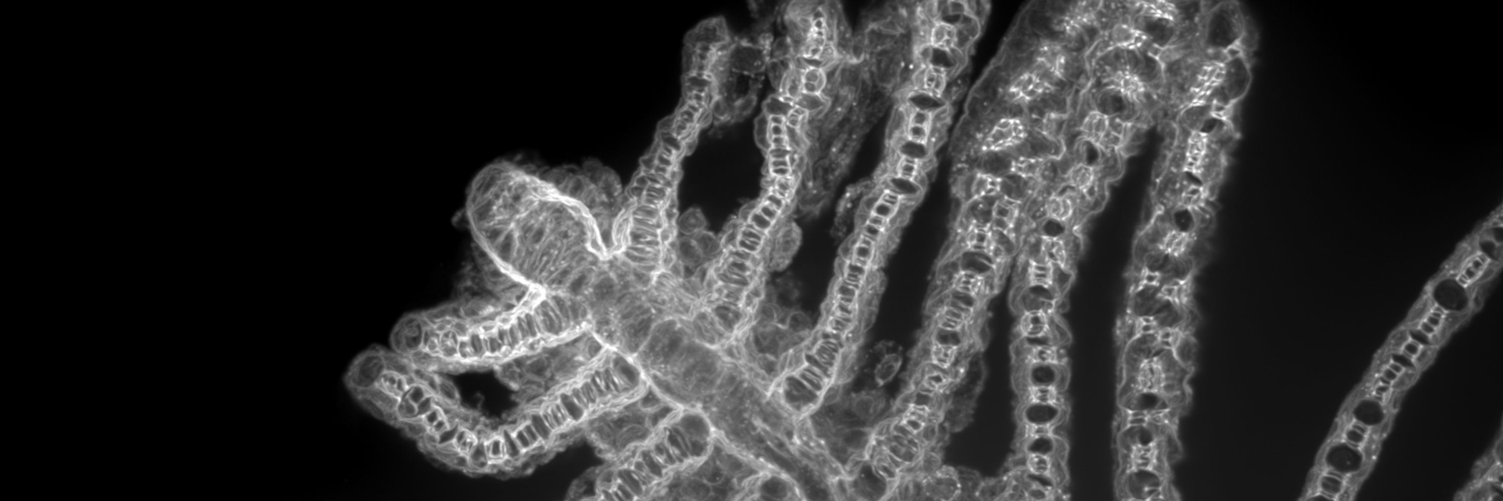
2/2
Also got a close-up where the expression pattern goes full-on pointillism
Among others, fgf10b is expressed in gill pillar cells, that structure the vascular network within the lamella.
Also got a close-up where the expression pattern goes full-on pointillism
Among others, fgf10b is expressed in gill pillar cells, that structure the vascular network within the lamella.
July 11, 2025 at 12:45 PM
2/2
Also got a close-up where the expression pattern goes full-on pointillism
Among others, fgf10b is expressed in gill pillar cells, that structure the vascular network within the lamella.
Also got a close-up where the expression pattern goes full-on pointillism
Among others, fgf10b is expressed in gill pillar cells, that structure the vascular network within the lamella.
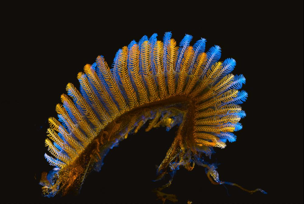
1/2
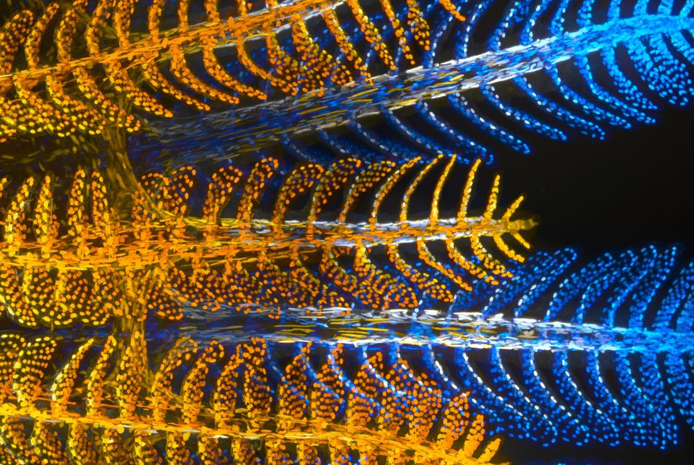
🧬🎨 When gills do Pointillism!
For this #FluorescenceFriday,
here’s a dissected gill arch expressing nuclear fgf10b:nEOS — depth color-coded: blue for medial filaments, yellow for lateral ones.
#Zebrafish #DevBio #Microscopy #Gills
🧬🎨 When gills do Pointillism!
For this #FluorescenceFriday,
here’s a dissected gill arch expressing nuclear fgf10b:nEOS — depth color-coded: blue for medial filaments, yellow for lateral ones.
#Zebrafish #DevBio #Microscopy #Gills
July 11, 2025 at 12:45 PM
1/2
🧬🎨 When gills do Pointillism!
For this #FluorescenceFriday,
here’s a dissected gill arch expressing nuclear fgf10b:nEOS — depth color-coded: blue for medial filaments, yellow for lateral ones.
#Zebrafish #DevBio #Microscopy #Gills
🧬🎨 When gills do Pointillism!
For this #FluorescenceFriday,
here’s a dissected gill arch expressing nuclear fgf10b:nEOS — depth color-coded: blue for medial filaments, yellow for lateral ones.
#Zebrafish #DevBio #Microscopy #Gills