


Dagan Segal
@dagansegal.bsky.social
quantitative imaging in pediatric cancer. postdoc @Danuser Lab, UTSW Find me at dagansegal.bsky.social
9/n This led us to a model where:
✳️ caveolae offer stable docking points for active PI3K to spread, inducing survival signaling in CD99 High cells.
✳️CD99 low cells don't have caveolae and must find other ways to regulate their signaling, leaving them at a disadvantage in certain contexts.
✳️ caveolae offer stable docking points for active PI3K to spread, inducing survival signaling in CD99 High cells.
✳️CD99 low cells don't have caveolae and must find other ways to regulate their signaling, leaving them at a disadvantage in certain contexts.

January 28, 2025 at 9:36 PM
9/n This led us to a model where:
✳️ caveolae offer stable docking points for active PI3K to spread, inducing survival signaling in CD99 High cells.
✳️CD99 low cells don't have caveolae and must find other ways to regulate their signaling, leaving them at a disadvantage in certain contexts.
✳️ caveolae offer stable docking points for active PI3K to spread, inducing survival signaling in CD99 High cells.
✳️CD99 low cells don't have caveolae and must find other ways to regulate their signaling, leaving them at a disadvantage in certain contexts.
8/n Using tools developed for quantitative 3D analysis- we saw that CD99 High cells had uniform active PI3K organization, which was lost upon knockdown of Caveolin-1!!

January 28, 2025 at 9:36 PM
8/n Using tools developed for quantitative 3D analysis- we saw that CD99 High cells had uniform active PI3K organization, which was lost upon knockdown of Caveolin-1!!
6/n- The CD99 High cells also
✳️ look CRAAZZY in fish xenografts👇
✳️show survival advantages in fish and mice
✳️show Caveolin-1 dependent survival advantage in response to chemotherapeutic drug (check out preprint for bullets 2 and 3!)
✳️ look CRAAZZY in fish xenografts👇
✳️show survival advantages in fish and mice
✳️show Caveolin-1 dependent survival advantage in response to chemotherapeutic drug (check out preprint for bullets 2 and 3!)

January 28, 2025 at 9:36 PM
6/n- The CD99 High cells also
✳️ look CRAAZZY in fish xenografts👇
✳️show survival advantages in fish and mice
✳️show Caveolin-1 dependent survival advantage in response to chemotherapeutic drug (check out preprint for bullets 2 and 3!)
✳️ look CRAAZZY in fish xenografts👇
✳️show survival advantages in fish and mice
✳️show Caveolin-1 dependent survival advantage in response to chemotherapeutic drug (check out preprint for bullets 2 and 3!)
5/n- second observation: these cells look really different also under and electron microscope!
The CD99 High cells have a ton of caveolae (e.g. "little caves" along the membrane) and higher expression of the structural caveolar component Caveolin-1 .
The CD99 High cells have a ton of caveolae (e.g. "little caves" along the membrane) and higher expression of the structural caveolar component Caveolin-1 .

January 28, 2025 at 9:36 PM
5/n- second observation: these cells look really different also under and electron microscope!
The CD99 High cells have a ton of caveolae (e.g. "little caves" along the membrane) and higher expression of the structural caveolar component Caveolin-1 .
The CD99 High cells have a ton of caveolae (e.g. "little caves" along the membrane) and higher expression of the structural caveolar component Caveolin-1 .
4/n - And also really distinct by flow cytometry targeting CD99- which means we can use flow to isolate these cells and do some cell biology!
First observation 👀: these cells look really different under a microscope!
First observation 👀: these cells look really different under a microscope!

January 28, 2025 at 9:36 PM
4/n - And also really distinct by flow cytometry targeting CD99- which means we can use flow to isolate these cells and do some cell biology!
First observation 👀: these cells look really different under a microscope!
First observation 👀: these cells look really different under a microscope!
3/n Our study started with the observation that when we passage Ewing Sarcoma cells non-enzymatically, a distinct population of cells emerges, with high expression of the glycoprotein and Ewing Sarcoma diagnostic marker CD99. Seen here by single cell transcriptomics-

January 28, 2025 at 9:36 PM
3/n Our study started with the observation that when we passage Ewing Sarcoma cells non-enzymatically, a distinct population of cells emerges, with high expression of the glycoprotein and Ewing Sarcoma diagnostic marker CD99. Seen here by single cell transcriptomics-

Thing 4 to gain attention and followers: Following the dynamic shape changes of a cancer cell over time.
December 10, 2024 at 1:30 AM
Thing 4 to gain attention and followers: Following the dynamic shape changes of a cancer cell over time.
Thing 3 to gain attention and followers: This was #NotTheCover of JCB - 3D rendering of a bunch of cancer cells imaged in zebrafish, pseudocolored by morphotype (e.g. shape classification)
December 10, 2024 at 1:30 AM
Thing 3 to gain attention and followers: This was #NotTheCover of JCB - 3D rendering of a bunch of cancer cells imaged in zebrafish, pseudocolored by morphotype (e.g. shape classification)
Thing 2 to gain attention and followers- cancer cells expressing an actin marker xenografted into zebrafish hindbrain ventricle tend to form these crazy pseudorosette-like structures. See Segal et al. JCB 2022
December 10, 2024 at 1:30 AM
Thing 2 to gain attention and followers- cancer cells expressing an actin marker xenografted into zebrafish hindbrain ventricle tend to form these crazy pseudorosette-like structures. See Segal et al. JCB 2022
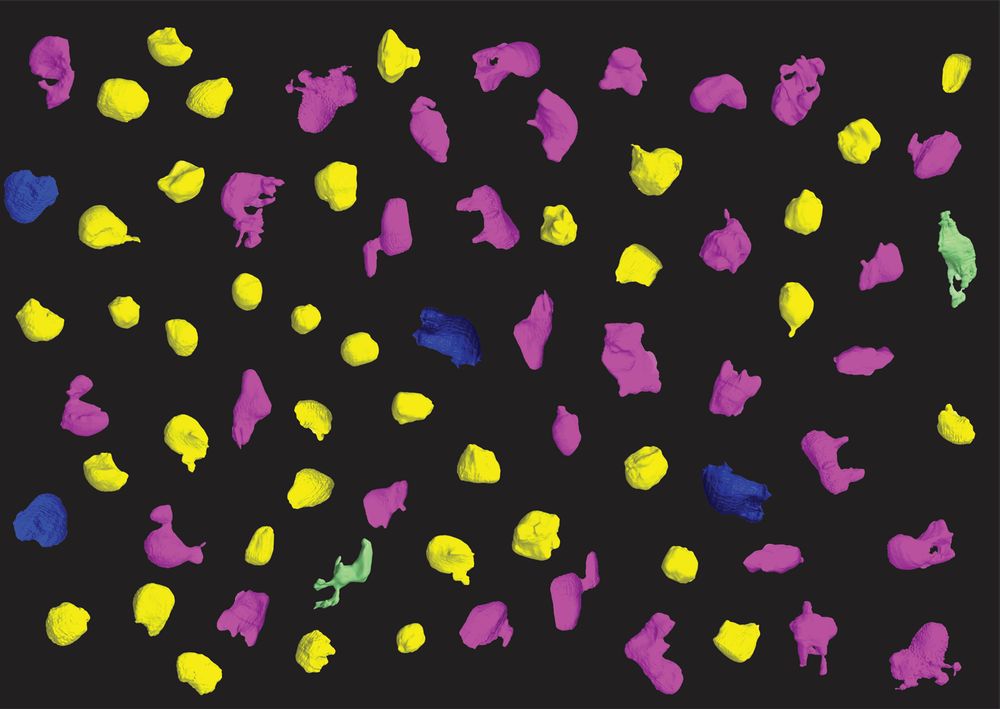
Sharing some random things I found on my desktop to gain followers:
Thing 1 - segmentation of a zebrafish xenografted with human cancer cells. Red is cancer cells, green is cell death marker, blue is proliferation marker.
Thing 1 - segmentation of a zebrafish xenografted with human cancer cells. Red is cancer cells, green is cell death marker, blue is proliferation marker.
December 10, 2024 at 1:30 AM
Sharing some random things I found on my desktop to gain followers:
Thing 1 - segmentation of a zebrafish xenografted with human cancer cells. Red is cancer cells, green is cell death marker, blue is proliferation marker.
Thing 1 - segmentation of a zebrafish xenografted with human cancer cells. Red is cancer cells, green is cell death marker, blue is proliferation marker.