


Saber C. Saharkhiz🔬📊💻🧬
@sabersaharkhiz.bsky.social
PhD student in Neuroscience program 🧠 (Dept. of CMM), The Ottawa Hospital, Faculty of Medicine, University of Ottawa ★ Molecular, Cell & Developmental Biologist ★ Quantitative Microscopist ★ Bioimage Analyst | DVM⚕️. Views are my own
Pinned

Excited to share the 1st part of my PhD work in @plosone.org ! 🚀
Progress in biology needs not only new ideas but also #workflow_automation
Open Access:
📃paper: journals.plos.org/plosone/arti...
💻Code: github.com/C-elegans-VN...
#CellBiology #Neuroscience #DeepLearning #Bioimage_Analysis
Progress in biology needs not only new ideas but also #workflow_automation
Open Access:
📃paper: journals.plos.org/plosone/arti...
💻Code: github.com/C-elegans-VN...
#CellBiology #Neuroscience #DeepLearning #Bioimage_Analysis
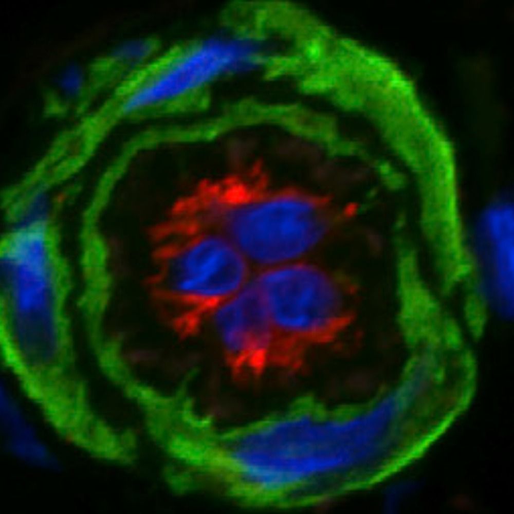
Do you know how many genetic tools used to visualize only the nuclei of these neurons? (From my PhD work)
1. Endogenous CRISPR knock-in tag
2. Promotor transgene fused to Histone
3. Endogenous CRISPR knock-in dual color tag with self-cleaving peptide T2A fused to Histone.
#FluorescenceFriday
1. Endogenous CRISPR knock-in tag
2. Promotor transgene fused to Histone
3. Endogenous CRISPR knock-in dual color tag with self-cleaving peptide T2A fused to Histone.
#FluorescenceFriday

November 9, 2025 at 5:39 AM
Do you know how many genetic tools used to visualize only the nuclei of these neurons? (From my PhD work)
1. Endogenous CRISPR knock-in tag
2. Promotor transgene fused to Histone
3. Endogenous CRISPR knock-in dual color tag with self-cleaving peptide T2A fused to Histone.
#FluorescenceFriday
1. Endogenous CRISPR knock-in tag
2. Promotor transgene fused to Histone
3. Endogenous CRISPR knock-in dual color tag with self-cleaving peptide T2A fused to Histone.
#FluorescenceFriday
Reposted by Saber C. Saharkhiz🔬📊💻🧬
Beating iPSC-derived heart muscle cells videoed through a microscope. Alpha-actinin-2 is shown. #CellBiology
November 9, 2025 at 3:12 AM
Beating iPSC-derived heart muscle cells videoed through a microscope. Alpha-actinin-2 is shown. #CellBiology
Reposted by Saber C. Saharkhiz🔬📊💻🧬
Required reading for cell biologists to get a sense of basic statistical principles!
www.nature.com/articles/s41...
www.nature.com/articles/s41...
Ten essential tips for robust statistics in cell biology - Nature Cell Biology
Statistical thinking is a core part of solid, trustworthy biology. However, many studies still include insufficient sample sizes, have poor experimental design or select an incorrect statistical metho...
www.nature.com
November 4, 2025 at 7:23 PM
Required reading for cell biologists to get a sense of basic statistical principles!
www.nature.com/articles/s41...
www.nature.com/articles/s41...
Reposted by Saber C. Saharkhiz🔬📊💻🧬
It's Halloween 🎃 #FlourescenceFriday! Here is a microscopic pumpkin generated by showing a cell nucleus in 🟠 with the primary cilium "stem" in 🟢! I hope everyone has a spooky 👻 day! 🔬 by @stjuderesearch.bsky.social grad student Christina Wang. 🧪

October 31, 2025 at 3:14 PM
It's Halloween 🎃 #FlourescenceFriday! Here is a microscopic pumpkin generated by showing a cell nucleus in 🟠 with the primary cilium "stem" in 🟢! I hope everyone has a spooky 👻 day! 🔬 by @stjuderesearch.bsky.social grad student Christina Wang. 🧪
Reposted by Saber C. Saharkhiz🔬📊💻🧬
#Atherosclerosis Epitranscriptomics
Mettl3-YTHDF3⏫m6A-MTRFA mRNA in SMCs➡️
⏫cholesterol-induced Mφ-like phenotype
SMC Mettl3 KO🐭+HFD AAV-PCSK9➡️
⏬Lesion burden, necrotic core, Mφ
👉Abolishes SMC contribution to lesion!
👉Reverted by AAV9-SM22-MRTFA
#CardiovascRes 2025
academic.oup.com/cardiovascre...
Mettl3-YTHDF3⏫m6A-MTRFA mRNA in SMCs➡️
⏫cholesterol-induced Mφ-like phenotype
SMC Mettl3 KO🐭+HFD AAV-PCSK9➡️
⏬Lesion burden, necrotic core, Mφ
👉Abolishes SMC contribution to lesion!
👉Reverted by AAV9-SM22-MRTFA
#CardiovascRes 2025
academic.oup.com/cardiovascre...

October 30, 2025 at 8:23 PM
#Atherosclerosis Epitranscriptomics
Mettl3-YTHDF3⏫m6A-MTRFA mRNA in SMCs➡️
⏫cholesterol-induced Mφ-like phenotype
SMC Mettl3 KO🐭+HFD AAV-PCSK9➡️
⏬Lesion burden, necrotic core, Mφ
👉Abolishes SMC contribution to lesion!
👉Reverted by AAV9-SM22-MRTFA
#CardiovascRes 2025
academic.oup.com/cardiovascre...
Mettl3-YTHDF3⏫m6A-MTRFA mRNA in SMCs➡️
⏫cholesterol-induced Mφ-like phenotype
SMC Mettl3 KO🐭+HFD AAV-PCSK9➡️
⏬Lesion burden, necrotic core, Mφ
👉Abolishes SMC contribution to lesion!
👉Reverted by AAV9-SM22-MRTFA
#CardiovascRes 2025
academic.oup.com/cardiovascre...

October 29, 2025 at 7:07 PM
Reposted by Saber C. Saharkhiz🔬📊💻🧬

Mechanical coordination between anaphase A and B drives asymmetric chromosome segregation, say Ana Dias Maia Henriques, Julien Dumont, Gilliane Maton and colleagues @ijmonod.bsky.social: rupress.org/jcb/article/...
#Development #CellCycle #CellDivision #Celegans #Mitosis
#Development #CellCycle #CellDivision #Celegans #Mitosis

October 29, 2025 at 1:31 PM
Mechanical coordination between anaphase A and B drives asymmetric chromosome segregation, say Ana Dias Maia Henriques, Julien Dumont, Gilliane Maton and colleagues @ijmonod.bsky.social: rupress.org/jcb/article/...
#Development #CellCycle #CellDivision #Celegans #Mitosis
#Development #CellCycle #CellDivision #Celegans #Mitosis
Reposted by Saber C. Saharkhiz🔬📊💻🧬
Episode 17 - perhaps my favorite yet. How do you write a forum.image.sc post that will get you the answers you need without revealing information you don't want to tell? @erinweisbart.bsky.social and I go through what the experts need to know to help you. Post your sci q's today on forum.image.sc !
October 27, 2025 at 6:20 PM
Episode 17 - perhaps my favorite yet. How do you write a forum.image.sc post that will get you the answers you need without revealing information you don't want to tell? @erinweisbart.bsky.social and I go through what the experts need to know to help you. Post your sci q's today on forum.image.sc !
Reposted by Saber C. Saharkhiz🔬📊💻🧬
As spooky season is upon us 🎃, we have cells that look like spider webs for #FluorescenceFriday 🧪🔬

October 24, 2025 at 7:03 PM
As spooky season is upon us 🎃, we have cells that look like spider webs for #FluorescenceFriday 🧪🔬
Reposted by Saber C. Saharkhiz🔬📊💻🧬
🧪 🤩 🔬
An iPSC-cardiac myocyte (heart muscle cell) in cell culture photographed through a microscope by former Burnette lab graduate student, Dr. James Hayes. We are preparing cover submissions for his last two first author papers from the lab! Actin filaments are shown. #CellBiology

October 22, 2025 at 4:20 PM
🧪 🤩 🔬
Reposted by Saber C. Saharkhiz🔬📊💻🧬
💡Just presented our new paper at the @iccv.bsky.social BioImage Computing workshop: cubic: CUDA-accelerated 3D Bioimage Computing. We introduce a simple way to add GPU acceleration to scikit-image–based bioimage processing pipelines by swapping import statements. 🧵
1/6 #iccv2025
1/6 #iccv2025
October 23, 2025 at 6:01 PM
💡Just presented our new paper at the @iccv.bsky.social BioImage Computing workshop: cubic: CUDA-accelerated 3D Bioimage Computing. We introduce a simple way to add GPU acceleration to scikit-image–based bioimage processing pipelines by swapping import statements. 🧵
1/6 #iccv2025
1/6 #iccv2025
Reposted by Saber C. Saharkhiz🔬📊💻🧬
We just hit 10 workshops submitted, with almost two weeks to go! Help us get to 20 before the last-day frantic mad dash? Teach your favorite tool today!
Halfway to I2K is BACK, friends of all kinds! Last year, 650 people attended 30+ TOTALLY FREE image analysis workshops of all kinds, across many timezones.
If you make image analysis software and want to teach it, workshop submissions are open now! We'd love to have your tool highlighted.
If you make image analysis software and want to teach it, workshop submissions are open now! We'd love to have your tool highlighted.

#HappyFluorescenceFriday!
#microscopycommunity- want to learn open source image analysis or share your knowledge to help others? We’ve got a FREE virtual workshop Nov 17-19! Now accepting workshop session applications!
Learn more & sign up: buff.ly/esGIotD
#microscopycommunity- want to learn open source image analysis or share your knowledge to help others? We’ve got a FREE virtual workshop Nov 17-19! Now accepting workshop session applications!
Learn more & sign up: buff.ly/esGIotD
October 20, 2025 at 2:52 PM
We just hit 10 workshops submitted, with almost two weeks to go! Help us get to 20 before the last-day frantic mad dash? Teach your favorite tool today!
Reposted by Saber C. Saharkhiz🔬📊💻🧬
Happy #FluorescenceFriday! This is a maximum intensity projection of depth shaded actin (🌈) in a section from an E9.5 🐭 neural tube (NT). 🔬 by postdoc @christinaadaly.bsky.social 👩🔬 🧪 Image shows the floor plate and lumen of the developing NT.
#SciArt #DevBio #DevNeuro
#SciArt #DevBio #DevNeuro

October 10, 2025 at 3:19 PM
Happy #FluorescenceFriday! This is a maximum intensity projection of depth shaded actin (🌈) in a section from an E9.5 🐭 neural tube (NT). 🔬 by postdoc @christinaadaly.bsky.social 👩🔬 🧪 Image shows the floor plate and lumen of the developing NT.
#SciArt #DevBio #DevNeuro
#SciArt #DevBio #DevNeuro
Reposted by Saber C. Saharkhiz🔬📊💻🧬

Today for #FluorescenceFriday I’m sharing a 👻Halloween-themed🎃 neural crest explant “web of cells” from Julia Godinez, a 4th year @ucdavis.bsky.social graduate student in the lab. She is studying mechanisms driving conserved and divergent cranial neural crest migration and differentiation. #DevBio

October 17, 2025 at 8:21 PM
Today for #FluorescenceFriday I’m sharing a 👻Halloween-themed🎃 neural crest explant “web of cells” from Julia Godinez, a 4th year @ucdavis.bsky.social graduate student in the lab. She is studying mechanisms driving conserved and divergent cranial neural crest migration and differentiation. #DevBio
Reposted by Saber C. Saharkhiz🔬📊💻🧬

Accurate somatic small variant discovery for multiple sequencing technologies with DeepSomatic www.nature.com/articles/s41... (read free: rdcu.be/eLny0) github.com/google/deeps...


October 16, 2025 at 7:29 PM
Accurate somatic small variant discovery for multiple sequencing technologies with DeepSomatic www.nature.com/articles/s41... (read free: rdcu.be/eLny0) github.com/google/deeps...

My son is so tired💤🤭😂🦜🪶

October 16, 2025 at 8:24 PM
My son is so tired💤🤭😂🦜🪶
Reposted by Saber C. Saharkhiz🔬📊💻🧬
Episode 16 - Learn more about the Cell Painting Gallery, a free repository of nearly a petabyte of images and imaging data, curated by the incredibly talented @erinweisbart.bsky.social . Truly a goldmine for big-data lovers everywhere!
October 14, 2025 at 11:48 AM
Episode 16 - Learn more about the Cell Painting Gallery, a free repository of nearly a petabyte of images and imaging data, curated by the incredibly talented @erinweisbart.bsky.social . Truly a goldmine for big-data lovers everywhere!
Reposted by Saber C. Saharkhiz🔬📊💻🧬
15 years in the making, we confirmed that mitochondria - the powerhouse of the cell - have an unusual localization in patients who experience psychosis (including schizophrenia and bipolar disorders). You’ll never guess what kind of patient cells we used to make this discovery… 🧵

October 10, 2025 at 4:47 PM
15 years in the making, we confirmed that mitochondria - the powerhouse of the cell - have an unusual localization in patients who experience psychosis (including schizophrenia and bipolar disorders). You’ll never guess what kind of patient cells we used to make this discovery… 🧵
Reposted by Saber C. Saharkhiz🔬📊💻🧬

Segment large images without tiling artifacts: sharing our work that should have been presented at ICCV in 2 weeks - the brilliant first author Elena can’t go because of visa issues.
The paper: arxiv.org/abs/2503.19545 1/🧵
The paper: arxiv.org/abs/2503.19545 1/🧵

October 9, 2025 at 12:56 PM
Segment large images without tiling artifacts: sharing our work that should have been presented at ICCV in 2 weeks - the brilliant first author Elena can’t go because of visa issues.
The paper: arxiv.org/abs/2503.19545 1/🧵
The paper: arxiv.org/abs/2503.19545 1/🧵
Reposted by Saber C. Saharkhiz🔬📊💻🧬

✨AI unveils hidden cellular worlds!🔬 University of Tsukuba's DeMemSeg precisely maps overlapping cell membranes in 2D images, unlocking disease insights! 🧬 #microscopy
Source: https://phys.org/news/2025-10-ai-based-method-accurately-segments.html
Source: https://phys.org/news/2025-10-ai-based-method-accurately-segments.html

AI-based method accurately segments and quantifies overlapping cell membranes
Researchers at University of Tsukuba have developed DeMemSeg, an AI-powered analysis pipeline that addresses a long-standing challenge in microscopy: precisely segmenting and measuring individual cell membranes that overlap in two-dimensional (2D) projection images. This innovation is expected to accelerate research on cellular mechanisms and related diseases.
phys.org
October 8, 2025 at 2:06 PM
✨AI unveils hidden cellular worlds!🔬 University of Tsukuba's DeMemSeg precisely maps overlapping cell membranes in 2D images, unlocking disease insights! 🧬 #microscopy
Source: https://phys.org/news/2025-10-ai-based-method-accurately-segments.html
Source: https://phys.org/news/2025-10-ai-based-method-accurately-segments.html
Just reached a small but meaningful milestone, 100+ citations in under 3 years since starting grad school. Grateful that my workare being recognized, and reminded of why I began this journey back at #Université_de_Sherbrooke and continued at the #University_of_Ottawa !

October 7, 2025 at 8:35 PM
Just reached a small but meaningful milestone, 100+ citations in under 3 years since starting grad school. Grateful that my workare being recognized, and reminded of why I began this journey back at #Université_de_Sherbrooke and continued at the #University_of_Ottawa !
Reposted by Saber C. Saharkhiz🔬📊💻🧬
My entry for today’s #FluorescenceFriday: a pupal #Drosophila testis with muscles expressing
🔵 lifeact &
🔴 RFP-nls
Honored & grateful to receive an honorable mention at @healthcare.nikon.com Nikon Small World 🌍🔬✨
🔗 www.nikonsmallworld.com/galleries/20...
#NikonSmallWorld #Microscopy #ScienceArt
🔵 lifeact &
🔴 RFP-nls
Honored & grateful to receive an honorable mention at @healthcare.nikon.com Nikon Small World 🌍🔬✨
🔗 www.nikonsmallworld.com/galleries/20...
#NikonSmallWorld #Microscopy #ScienceArt
October 3, 2025 at 9:06 AM
My entry for today’s #FluorescenceFriday: a pupal #Drosophila testis with muscles expressing
🔵 lifeact &
🔴 RFP-nls
Honored & grateful to receive an honorable mention at @healthcare.nikon.com Nikon Small World 🌍🔬✨
🔗 www.nikonsmallworld.com/galleries/20...
#NikonSmallWorld #Microscopy #ScienceArt
🔵 lifeact &
🔴 RFP-nls
Honored & grateful to receive an honorable mention at @healthcare.nikon.com Nikon Small World 🌍🔬✨
🔗 www.nikonsmallworld.com/galleries/20...
#NikonSmallWorld #Microscopy #ScienceArt
Reposted by Saber C. Saharkhiz🔬📊💻🧬

💫NEW: @telemanlab.bsky.social & co demonstrate that #AMPK can be activated by signaling metabolite, #adenosine, under non-stress conditions during #Drosophila development. The intestine regulates adenosine levels, thus, remotely controlling wing disc AMPK activation and growth.
bit.ly/4gX6njF
bit.ly/4gX6njF

Remote control of AMPK via extracellular adenosine controls tissue growth - Nature Cell Biology
Zhang et al. demonstrate that AMPK can be activated by signalling metabolite, adenosine, under non-stress conditions during Drosophila development. The intestine regulates adenosine levels, thus, remotely controlling wing disc AMPK activation and growth.
bit.ly
October 4, 2025 at 2:32 PM
💫NEW: @telemanlab.bsky.social & co demonstrate that #AMPK can be activated by signaling metabolite, #adenosine, under non-stress conditions during #Drosophila development. The intestine regulates adenosine levels, thus, remotely controlling wing disc AMPK activation and growth.
bit.ly/4gX6njF
bit.ly/4gX6njF
Reposted by Saber C. Saharkhiz🔬📊💻🧬

#fluorescencefriday special. Half marmoset 🧠 cleared and stained using SHIELD and imaged by lightsheet microscopy. Courtesy of Jesleen Saini @shahrzadbahrampour.bsky.social and @stefan-everling.bsky.social
October 3, 2025 at 4:00 PM
#fluorescencefriday special. Half marmoset 🧠 cleared and stained using SHIELD and imaged by lightsheet microscopy. Courtesy of Jesleen Saini @shahrzadbahrampour.bsky.social and @stefan-everling.bsky.social