Joyce Meiring
@joycemeiri.bsky.social
730 followers 180 following 17 posts
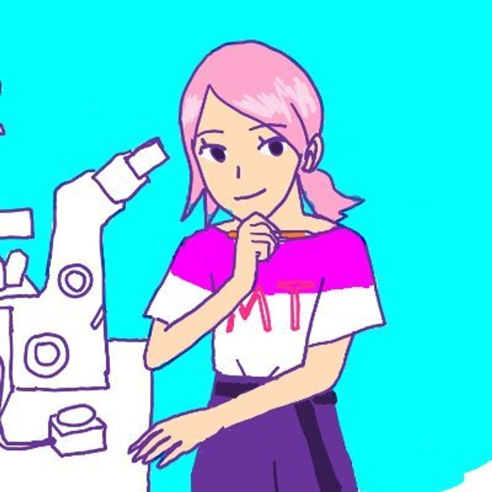
Postdoc in Akhmanova lab, Utrecht University. Cytoskeleton, 3D migration & light switchable cell biology tools | she/her
Posts Media Videos Starter Packs
Pinned
Hi! My name is Joyce and I love building and using tools to manipulate the #cytoskeleton of cells to better understand their function. 🔬
Below you can see my movie of #opto-katanin ✂️(SspB-mCh-p60) being used to locally disassemble #microtubules (labelled by SiR-tubulin) inside a neuron.
Reposted by Joyce Meiring
A cell finding its way through the matrix, imaged with @joycemeiri.bsky.social on LLS.
Reposted by Joyce Meiring
Our new preprint is about the airway epithelium microtubule network, cilia, basal body protein composition, averaging of volumetric fluorescence data, and expansion microscopy.
Four years of very hard work from our very talented Emma van Grinsven. www.biorxiv.org/content/10.1... (1/N)
Reposted by Joyce Meiring
Our September issue is here! rupress.org/jcb/issue/22...
Airyscan super-resolution immunofluorescence image of HeLa cells stained for two types of intermediate filaments: vimentin (magenta) & keratin-8 (cyan). From Pasolli, @joycemeiri.bsky.social, Akhmanova et al. rupress.org/jcb/article/...
All the opto-vimentin plasmids are now available on Addgene: www.addgene.org/Anna_Akhmano...
Radial microtubules in cyan and non-radial microtubules in yellow.
Biochemical tools to rapidly manipulate vimentin intermediate filaments by means of chemicals or light (optogenetics)

📷 Milena Pasolli & @joycemeiri.bsky.social et al
‪@utrechtuniversity.bsky.social in @jcb.org

➡️ bpod.org.uk/archive/2025...
Reposted by Joyce Meiring
Optogenetic and chemical genetic tools for rapid repositioning of #vimentin intermediate filaments. New tools developed by Milena Pasolli, Joyce Meiring @joycemeiri.bsky.social, Anna Akhmanova and colleagues @utrechtuniversity.bsky.social: rupress.org/jcb/article/...

@gijsjekoenderink.bsky.social
Reposted by Joyce Meiring
New: Pasolli, Meiring, Akhmanova et al. @utrechtuniversity.bsky.social describe tools to acutely relocate #vimentin intermediate filaments by inducibly coupling them to microtubule motors. rupress.org/jcb/article/...

@joycemeiri.bsky.social @gijsjekoenderink.bsky.social
#Technology #Cytoskeleton
Here is an example of the reversibility. The first 30 min the cell is pulsed with blue light (when blue dot is shown) causing the tagged vimentin to associate with - end oriented kinesins that pull the vimentin to the cell centre. Then blue light pulsing is stopped and vimentin redistributes.
Excited to share that our fast & reversible chemical and optogenetic vimentin pulling tools are now published in JCB! 🥳
We show a subset of organelles are displaced with vimentin pulling to the cell centre and cells become much softer despite little or no effect on actin/MTs
doi.org/10.1083/jcb....
Reposted by Joyce Meiring
Thrilled to share our latest preprint from @yiechanglin.bsky.social & @ciarawallis.bsky.social showing that AF2-Multimer and AF3 can quickly & accurately predict the oligomeric states & structures of proteins (& maybe can identify multiple biologically relevant oligomeric states 👀)
tiny.cc/oligomers
Don't forget to register, abstract submission deadline is next week March 14. 😉
Come join us in Utrecht for some cutting edge #microscopy and 3D cell biology!! #3Dcellbio
Congratulations!! 🥳🥂
Is this an animated Jackson Pollock painting or live cell tracking movie? 😍
Come join us in Utrecht for some cutting edge #microscopy and 3D cell biology!! #3Dcellbio
Hi! I was wondering if anyone had successfully cultured happy HEK293T cells in defined serum alternatives? If so, what did you use?
Excited to share our new pre-print showcasing tools (chemical & optogenetic) for repositioning Vimentin in cells! Movie shows ER (Halo-KDEL) sheets being relocalized together with vimentin (Vim) upon local vimentin pulling in U2OS. @gijsjekoenderink.bsky.social
www.biorxiv.org/content/10.1...
Reposted by Joyce Meiring
Time to shine for intermediate filaments! An optogenetic vimentin perturbation strategy reveals its role for ER and mitochondria positioning (but no effect on actin or microtubules patterns) @joycemeiri.bsky.social
www.biorxiv.org/content/10.1...
CAMSAP3 stretches (microtubule -end binding protein) are shown in yellow. They move towards the Soma in developing neurons.
Hi! I'm Max. I'm developing a #biophysical model of #microtubules in #neurons during #development. The model is based on my surprising experimental finding (lots of live-cell imaging) that the entire microtubule array in neurites flows retrogradely into the soma: www.science.org/doi/10.1126/... 🧪 🧠📈
We like the Cytochrome C from BD Pharmingen cat#556432
This video shows the smallest area I've managed to sever inside a U2OS cell. The goal for this experiment was to sever microtubules around a subset of the Golgi (labelled here in cyan, together with exocytic vesicles) to see what would happen to it's morphology/localization.
However in a neuron it could be a smaller area still, because diffusion is slower in neurites due to their morphology. I haven't tried to see how small the area could be in neurons though.
Good question, I would estimate an area of around 10 um^2.
Hi! My name is Joyce and I love building and using tools to manipulate the #cytoskeleton of cells to better understand their function. 🔬
Below you can see my movie of #opto-katanin ✂️(SspB-mCh-p60) being used to locally disassemble #microtubules (labelled by SiR-tubulin) inside a neuron.