Rachael Ott
@rachaelott.bsky.social
2.4K followers 2.4K following 160 posts
Neurobiologist @UofSC 🔬 Super-resolution #microscopy, synaptic plasticity, cytoskeleton, #neurodegeneration 🦠
Posts Media Videos Starter Packs
Pinned
rachaelott.bsky.social
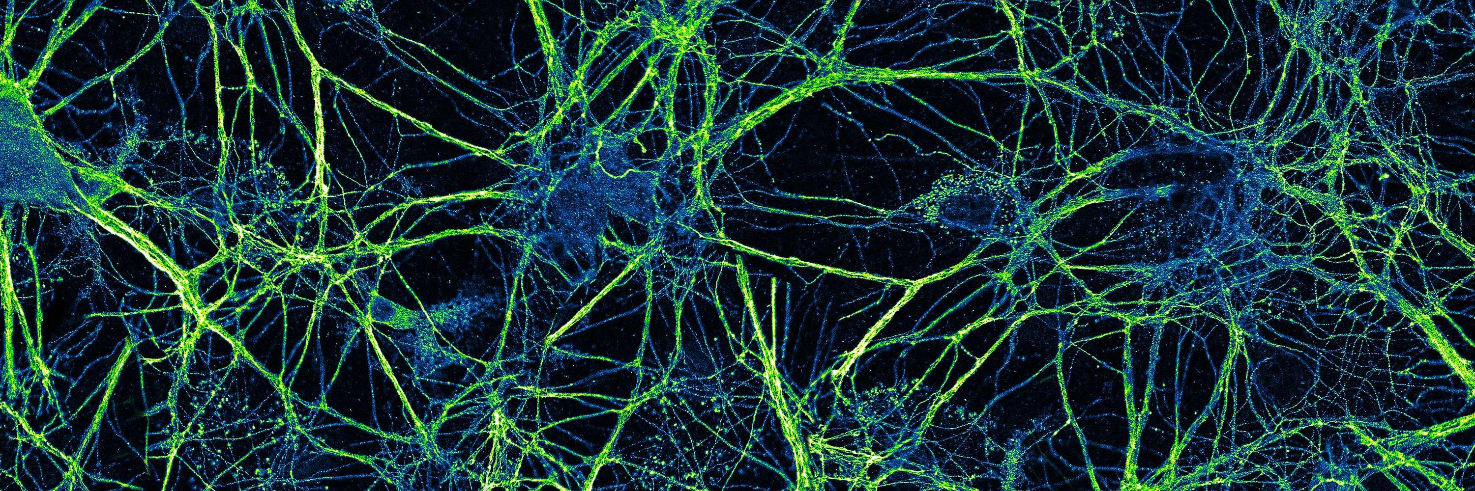
For #FluorescenceFriday, tau (blue) and long loops of neurofilament proteins are shown in a primary hippocampal neuron 🔬 #Neuroscience #Microscopy
Tau (blue) and neurofilament (fire) are shown in a primary hippocampal neuron.
Reposted by Rachael Ott
kaimurk.bsky.social
This #microscopymonday is a cleaning day. Watch how a #microglia cell mops up and phagozytoses debris and junk in a very messy neuronal co-culture. BTW, it was a proper cleaning job → the neurons made it and formed nice networks
Phase contrast imaging started at DIV1. One frame per 20 min.
Reposted by Rachael Ott
mag2art.bsky.social
Fish keratocytes isolated from a fish scale crawling around. Technique: DIC microscopy. #CellBiology.
Another #NikonSmallWorldReject
Reposted by Rachael Ott
rachaelott.bsky.social
For #FluorescenceFriday, α-spectrin (red) and lipid droplets (green) are shown in adipocytes 🔬 #Microscopy #CellBiology
α-spectrin (red), lipid droplets (green), and nuclei (blue) are shown in adipocytes. Scale bar is 20 μm.
rachaelott.bsky.social
Thank you! It’s BODIPY 505/510
rachaelott.bsky.social
For #FluorescenceFriday, α-spectrin (red) and lipid droplets (green) are shown in adipocytes 🔬 #Microscopy #CellBiology
α-spectrin (red), lipid droplets (green), and nuclei (blue) are shown in adipocytes. Scale bar is 20 μm.
Reposted by Rachael Ott
daniellebeckman.bsky.social
Viral trigger in Alzheimer’s disease is the real scary spooky #FluorescenceFriday.
Amyloid-β🟣 is an antimicrobial peptide that can be produced to trap pathogens in the brain. But it also induces an extensive neuroinflammatory response, as shown here with 🟠astrocytes. #NeuroCovid
Confocal microscopy photo showing astrocytes and amyloid-B
Reposted by Rachael Ott
haoyin.bsky.social
Sometimes, poor perfusion is attributed to a Venule🤓

Age-related rarefaction of capillary meshwork converging into principal cortical venules, most pronounced in layer 6/CC➡️
Gliosis+demyelination in 🐭 #WhiteMatterAging

#SciArt
@andyshihlab.bsky.social #NatNeuro 2025
www.nature.com/articles/s41...
Reposted by Rachael Ott
andreacaldarola.bsky.social
For #FluorescenceFriday : 3 cells start at the same Ca2+ concentration but undergo 3 different Histamine responses. Imaged using the lifetime-based sensor Tq-Ca-FLITS.
Reposted by Rachael Ott
christlet.bsky.social
Beautiful live-cell imaging of lysosome tubulation along microtubules in this work just out from @lysoluis.bsky.social et al:
rupress.org/jcb/article/...
Reposted by Rachael Ott
schalleklab.bsky.social
#FluorescenceFriday
Watch to the end! Immune cells in the retina remain flat until damage is detected. Cells change their shape and dive deep in response to damage (pink cell)
@cvsuor.bsky.social @urochester.bsky.social
@flaumeye.bsky.social @uofrbme.bsky.social
elifesciences.org/articles/98662
Reposted by Rachael Ott
labogden.bsky.social
Happy #FluorescenceFriday! This is a maximum intensity projection of depth shaded actin (🌈) in a section from an E9.5 🐭 neural tube (NT). 🔬 by postdoc @christinaadaly.bsky.social 👩‍🔬 🧪 Image shows the floor plate and lumen of the developing NT.

#SciArt #DevBio #DevNeuro
Rainbow shading of a confocal micrograph of a developing mouse neural tube. The protein actin is stained and colored in rainbow to indicate depth in the section.
Reposted by Rachael Ott
natclarke.bsky.social
🚨 My lab is hiring at all levels!

Interested in animal origins & evolutionary cell biology?

I'm recruiting a postdoc, PhD students & a research assistant to study the molecular evolution of cell adhesion using marine invertebrates + comparative genomics.

🔗: clarkelab.com/join/

Please repost!
Reposted by Rachael Ott
jcb.org
Geng, @kjverhey1.bsky.social et al find that KIF1C forms condensates where the exposed #kinesin motor domains entangle nearby #microtubules, causing them to bend & break in a process dependent on motor processivity, cluster properties, cytoplasmic viscosity, & MT anchors rupress.org/jcb/article/...
Reposted by Rachael Ott
manuelthery.bsky.social
What would happen if cells would express more molecular motors ?
They would break all microtubules 😱
As shown by recent great work from @kjverhey1.bsky.social

rupress.org/jcb/article-...
Reposted by Rachael Ott
retof.bsky.social
We present multi-immersion Oblique Plane microscope (miOPM), a light-sheet platform that can be adapted to a wide range of applications, from sensitive live cell imaging to imaging organs and cleared tissues.
www.biorxiv.org/content/10.1...
Reposted by Rachael Ott
bucklingmt.bsky.social
Playing around with @kwolbachia.bsky.social 's LUT manager plugin. Absolutely love the campfire LUT🔥 😍#microtubulemonday
Reposted by Rachael Ott
socialinfluenza.bsky.social
This is incredibly beautiful, and the fact that the resolution is sufficient to follow the path of DNA spooling inside the virion head and through the tail is amazing
daumlab.bsky.social
Out in Science Advances: Our #cryoEM structure of HFTV1, a virus infecting the halophile #archaea. *First full atomic structure (containing all structural proteins) of any tailed virus!* Congrats and thanks to all co-authors and our fantastic collaborators! www.science.org/doi/10.1126/...
Cryo-EM resolves the structure of the archaeal dsDNA virus HFTV1 from head to tail
This structure of an archaeal tailed virus (arTV) provides detailed insights into arTV assembly and infection mechanisms.
www.science.org
Reposted by Rachael Ott
rachaelott.bsky.social
For #FluorescenceFriday, RhoA (blue) and the actin cytoskeleton (magenta) are shown in a set of primary microglia 🔬 #Neuroscience #Microscopy
RhoA (blue) and the actin cytoskeleton (magenta) are shown in a set of primary microglia.
rachaelott.bsky.social
Thank you Shweta! 😄
Reposted by Rachael Ott
llobrienlab.bsky.social
Sharing this excellent review now out from postdoc Sarah McLarnon! Lots of useful info on 3D imaging, quantification of imaging data, and how this has been applied to the kidney. And since it's #FluorescenceFriday, sharing a figure with our own imaging data!
www.sciencedirect.com/science/arti...
Figure from our review paper showing various fluorescence microscopy images of cells and structures of the kidney