


Santosh Vardhana
@santoshvardhana.bsky.social
Researching immunology, metabolism, and cancer, and caring for patients with lymphoma at MSKCC (www.vardhanalab.com). Opinions are my own.

How can this be real? It is - this is a very real post by the current acting director of the CDC - essentially a full confession of scientific illiteracy. God help us.

September 4, 2025 at 11:09 PM
How can this be real? It is - this is a very real post by the current acting director of the CDC - essentially a full confession of scientific illiteracy. God help us.
Sequencing of nascently transcribed RNAs during chronic TCR stimulation revealed that chronic MEK activation drives preferential transcription of genes expressed by terminally exhausted T cells, including immune checkpoints but also cytotoxic genes.

May 14, 2025 at 5:06 PM
Sequencing of nascently transcribed RNAs during chronic TCR stimulation revealed that chronic MEK activation drives preferential transcription of genes expressed by terminally exhausted T cells, including immune checkpoints but also cytotoxic genes.
Tanmana confirmed that persistent antigen encounter, both in vitro and in vivo, was sufficient to increase nascent transcription rates in a MEK dependent fashion.

May 14, 2025 at 5:06 PM
Tanmana confirmed that persistent antigen encounter, both in vitro and in vivo, was sufficient to increase nascent transcription rates in a MEK dependent fashion.
Indeed, acute cycloheximide treatment confirmed that the increased mitochondrial oxygen consumption rate in chronically stimulated T cells is driven by protein synthesis and reversed by MEK inhibition.

May 14, 2025 at 5:06 PM
Indeed, acute cycloheximide treatment confirmed that the increased mitochondrial oxygen consumption rate in chronically stimulated T cells is driven by protein synthesis and reversed by MEK inhibition.
Using click chemistry, Tanmana demonstrated that MEK inhibition attenuated the rate of protein translation, indicating that MEK sets the bioenergetic demand of exhausted T cells by altering the balance of ATP synthesis versus NAD+ regeneration.

May 14, 2025 at 5:06 PM
Using click chemistry, Tanmana demonstrated that MEK inhibition attenuated the rate of protein translation, indicating that MEK sets the bioenergetic demand of exhausted T cells by altering the balance of ATP synthesis versus NAD+ regeneration.
This is consistent with pioneering work from Martin Brand showing that in proliferating rat thymocytes, protein synthesis is a substantial (or even dominant) source of ATP demand.

May 14, 2025 at 5:06 PM
This is consistent with pioneering work from Martin Brand showing that in proliferating rat thymocytes, protein synthesis is a substantial (or even dominant) source of ATP demand.
Tanmana next asked how MEK regulates bioenergetic demand during chronic TCR stimulation. RNA-seq analysis led her to hypothesize that MEK regulates bioenergetic demand in Texh at the level of protein synthesis.

May 14, 2025 at 5:06 PM
Tanmana next asked how MEK regulates bioenergetic demand during chronic TCR stimulation. RNA-seq analysis led her to hypothesize that MEK regulates bioenergetic demand in Texh at the level of protein synthesis.
As a result, MEK inhibition restored T cell proliferation, particularly under conditions of limited nutrient availability, and improved TIL persistence while attenuating terminal T cell exhaustion.

May 14, 2025 at 5:06 PM
As a result, MEK inhibition restored T cell proliferation, particularly under conditions of limited nutrient availability, and improved TIL persistence while attenuating terminal T cell exhaustion.
Tanmana tested every proximal TCR signaling component and identified MEK as the primary driver of nutrient uptake, mitochondrial ATP production, and ROS accumulation; consequently, targeting MEK reduced nutrient uptake and rebalanced NADH/NAD+ ratios.

May 14, 2025 at 5:06 PM
Tanmana tested every proximal TCR signaling component and identified MEK as the primary driver of nutrient uptake, mitochondrial ATP production, and ROS accumulation; consequently, targeting MEK reduced nutrient uptake and rebalanced NADH/NAD+ ratios.
This paradigm, however, is broken during chronic TCR stimulation, which drives mitochondrial NADH accumulation to support ATP synthesis, ultimately leading to ROS generation and exhaustion.

May 14, 2025 at 5:06 PM
This paradigm, however, is broken during chronic TCR stimulation, which drives mitochondrial NADH accumulation to support ATP synthesis, ultimately leading to ROS generation and exhaustion.
She further showed that this rate of NADH generation was due primarily to increased rates of glucose uptake and entry of glucose-derived carbons into the TCA cycle.

May 14, 2025 at 5:06 PM
She further showed that this rate of NADH generation was due primarily to increased rates of glucose uptake and entry of glucose-derived carbons into the TCA cycle.
Tanmana found that signs of increased NADH/NAD+ were present immediately upon chronic TCR stimulation, despite unchanged rates of oxygen consumption compared to conventionaly stimulated cells, favoring excess NADH generation over ETC dysfunction as the primary source of ROS.

May 14, 2025 at 5:06 PM
Tanmana found that signs of increased NADH/NAD+ were present immediately upon chronic TCR stimulation, despite unchanged rates of oxygen consumption compared to conventionaly stimulated cells, favoring excess NADH generation over ETC dysfunction as the primary source of ROS.
First, electrons could be delivered to the ETC at a rate that exceeded the ability to deliver electrons to molecular oxygen. Alternatively, either lack of an electron acceptor (O2) or a defect in electron transport could lead to an accumulation of free electrons.

May 14, 2025 at 5:06 PM
First, electrons could be delivered to the ETC at a rate that exceeded the ability to deliver electrons to molecular oxygen. Alternatively, either lack of an electron acceptor (O2) or a defect in electron transport could lead to an accumulation of free electrons.
Specifically, buffering mitochondrial reactive oxygen species (ROS) with N-acetylcysteine was sufficient to block chronic TCR-driven loss of proliferation and terminal differentiation.

May 14, 2025 at 5:06 PM
Specifically, buffering mitochondrial reactive oxygen species (ROS) with N-acetylcysteine was sufficient to block chronic TCR-driven loss of proliferation and terminal differentiation.
In summary, we believe that suppression of intratumoral protein synthesis is a novel bottleneck on TIL function, including in patients, where a recently identified ‘stressed’ T cell population that predicts immunotherapy failure shows signs of substantial amino acid stress.

May 12, 2025 at 10:55 PM
In summary, we believe that suppression of intratumoral protein synthesis is a novel bottleneck on TIL function, including in patients, where a recently identified ‘stressed’ T cell population that predicts immunotherapy failure shows signs of substantial amino acid stress.
Kevin was able to show that either blocking glutamine anaplerosis via glutaminase inhibition or promoting glutamine-independent amino acid uptake by overexpressing the sodium-amino acid co-transporter SLC6A15 improved protein synthesis rates and anti-tumor immunity.

May 12, 2025 at 10:55 PM
Kevin was able to show that either blocking glutamine anaplerosis via glutaminase inhibition or promoting glutamine-independent amino acid uptake by overexpressing the sodium-amino acid co-transporter SLC6A15 improved protein synthesis rates and anti-tumor immunity.
It turns out that T cells utilize quite a bit of glutamine to a) replenish TCA cycle intermediates (‘anaplerosis’) as well as to b) exchange for other amino acids.

May 12, 2025 at 10:55 PM
It turns out that T cells utilize quite a bit of glutamine to a) replenish TCA cycle intermediates (‘anaplerosis’) as well as to b) exchange for other amino acids.
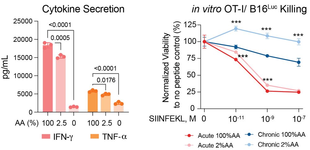
Answer #2: Unsurprisingly, amino acid-dependent protein synthetic capacity is a major regulator of the ability of T cells to release inflammatory cytokines and kill tumor cells.
May 12, 2025 at 10:55 PM
Answer #2: Unsurprisingly, amino acid-dependent protein synthetic capacity is a major regulator of the ability of T cells to release inflammatory cytokines and kill tumor cells.
Kevin adapted this reporter for T cells and showed that the reporter was activated by T cells, but not tumor cells, in vivo, despite the fact that amino acid deprivation activated the reporter in both cell types in vitro.

May 12, 2025 at 10:55 PM
Kevin adapted this reporter for T cells and showed that the reporter was activated by T cells, but not tumor cells, in vivo, despite the fact that amino acid deprivation activated the reporter in both cell types in vitro.
This functional defect is not a late or rare event in TILs - quite the opposite. Essentially all TILs exhibit decreased protein synthesis rates, and using adoptive transfer experiments Kevin showed that TILs lose protein synthesis within 24 hours of tumor infiltration.

May 12, 2025 at 10:55 PM
This functional defect is not a late or rare event in TILs - quite the opposite. Essentially all TILs exhibit decreased protein synthesis rates, and using adoptive transfer experiments Kevin showed that TILs lose protein synthesis within 24 hours of tumor infiltration.
Shockingly, he found that T cells were uniquely limited in their ability to synthesize proteins within tumors – other cells within tumors synthesized proteins at higher rates, as did T cells within draining lymph nodes.

May 12, 2025 at 10:55 PM
Shockingly, he found that T cells were uniquely limited in their ability to synthesize proteins within tumors – other cells within tumors synthesized proteins at higher rates, as did T cells within draining lymph nodes.
Kevin wondered if this disconnect might be due to a defect in the ability of T cells to translate mRNA within tumors. To test this hypothesis, he employed click chemistry to label nascently synthesized proteins within cells in vivo (hat tip
@carolynbertozzi.bskyverified.social)
@carolynbertozzi.bskyverified.social)

May 12, 2025 at 10:55 PM
Kevin wondered if this disconnect might be due to a defect in the ability of T cells to translate mRNA within tumors. To test this hypothesis, he employed click chemistry to label nascently synthesized proteins within cells in vivo (hat tip
@carolynbertozzi.bskyverified.social)
@carolynbertozzi.bskyverified.social)
Lack of response is therefore caused by either an inadequate reservoir of precursor cells or an inability to acquire effector function within tumors.

May 12, 2025 at 10:55 PM
Lack of response is therefore caused by either an inadequate reservoir of precursor cells or an inability to acquire effector function within tumors.
Moreover, to illustrate that LN disruption by chronic inflammation can be overcome, we provide a vignette of an exceptional responder to neoadjuvant therapy for GC whose LN converted phenotypes; this was associated with a dramatic improvement in intratumoral T cell surveillance.

May 10, 2025 at 11:49 PM
Moreover, to illustrate that LN disruption by chronic inflammation can be overcome, we provide a vignette of an exceptional responder to neoadjuvant therapy for GC whose LN converted phenotypes; this was associated with a dramatic improvement in intratumoral T cell surveillance.