


Polina Rusina
@polinarusina.bsky.social
PhD student at Biozentrum, Basel🇨🇭
Before: Moscow State University Neuroscience'20 :: Max Planck Florida Institute of Neuroscience'23 :: MS from Scheiffele lab
Before: Moscow State University Neuroscience'20 :: Max Planck Florida Institute of Neuroscience'23 :: MS from Scheiffele lab
Reposted by Polina Rusina

This #microscopymonday is a cleaning day. Watch how a #microglia cell mops up and phagozytoses debris and junk in a very messy neuronal co-culture. BTW, it was a proper cleaning job → the neurons made it and formed nice networks
Phase contrast imaging started at DIV1. One frame per 20 min.
Phase contrast imaging started at DIV1. One frame per 20 min.
October 12, 2025 at 4:32 PM
This #microscopymonday is a cleaning day. Watch how a #microglia cell mops up and phagozytoses debris and junk in a very messy neuronal co-culture. BTW, it was a proper cleaning job → the neurons made it and formed nice networks
Phase contrast imaging started at DIV1. One frame per 20 min.
Phase contrast imaging started at DIV1. One frame per 20 min.
Reposted by Polina Rusina
Integrated prediction of RNA secondary structure jointly with 3D motifs and pseudoknots guided by evolutionary information.
@aakaran31.bsky.social and @rivaselenarivas.bsky.social
link.springer.com/article/10.1...
@aakaran31.bsky.social and @rivaselenarivas.bsky.social
link.springer.com/article/10.1...

All-at-once RNA folding with 3D motif prediction framed by evolutionary information - Nature Methods
Structural RNAs exhibit a vast array of recurrent short three-dimensional (3D) elements found in loop regions involving non-Watson–Crick interactions that help arrange canonical double helices into tertiary structures. Here we present CaCoFold-R3D, a probabilistic grammar that predicts these RNA 3D motifs (also termed modules) jointly with RNA secondary structure over a sequence or alignment. CaCoFold-R3D uses evolutionary information present in an RNA alignment to reliably identify canonical helices (including pseudoknots) by covariation. Here we further introduce the R3D grammars, which also exploit helix covariation that constrains the positioning of the mostly noncovarying RNA 3D motifs. Our method runs predictions over an almost-exhaustive list of over 50 known RNA motifs (‘everything’). Motifs can appear in any nonhelical loop region (including three-way, four-way and higher junctions) (‘everywhere’). All structural motifs as well as the canonical helices are arranged into one single structure predicted by one single joint probabilistic grammar (‘all-at-once’). Our results demonstrate that CaCoFold-R3D is a valid alternative for predicting the all-residue interactions present in a RNA 3D structure. CaCoFold-R3D is fast and easily customizable for novel motif discovery and shows promising value both as a strong input for deep learning approaches to all-atom structure prediction as well as toward guiding RNA design as drug targets for therapeutic small molecules.
link.springer.com
October 3, 2025 at 12:42 PM
Integrated prediction of RNA secondary structure jointly with 3D motifs and pseudoknots guided by evolutionary information.
@aakaran31.bsky.social and @rivaselenarivas.bsky.social
link.springer.com/article/10.1...
@aakaran31.bsky.social and @rivaselenarivas.bsky.social
link.springer.com/article/10.1...
Reposted by Polina Rusina

Excited to highlight a new paper from my graduate student Sergio Bernal Garcia who, together withLuke Hammond, developed RESPAN, a new deep-learning pipeline automating the segmentation of neuronal morphology and dendritic spines from fluorescent microscopy images:
www.cell.com/cell-reports...
www.cell.com/cell-reports...

A deep learning pipeline for accurate and automated restoration, segmentation, and quantification of dendritic spines
Garcia et al. present RESPAN, a pipeline that enables automated dendritic spine analysis
through seamless integration of deep learning image restoration and segmentation capabilities
with comprehensiv...
www.cell.com
September 21, 2025 at 1:01 PM
Excited to highlight a new paper from my graduate student Sergio Bernal Garcia who, together withLuke Hammond, developed RESPAN, a new deep-learning pipeline automating the segmentation of neuronal morphology and dendritic spines from fluorescent microscopy images:
www.cell.com/cell-reports...
www.cell.com/cell-reports...
Reposted by Polina Rusina

🧠🦈Excited to present our latest work🧠🦈Interested in brain evolution? And shark embryos? Then read on… Our work sheds light on the deep origins of our brain’s most complex regions.
September 2, 2025 at 5:51 PM
🧠🦈Excited to present our latest work🧠🦈Interested in brain evolution? And shark embryos? Then read on… Our work sheds light on the deep origins of our brain’s most complex regions.
Reposted by Polina Rusina
A proteome-wide screen for membrane-interactions in intrinsically disordered regions of transmembrane proteins reveals a role in disease www.biorxiv.org/content/10.1...

A proteome-wide screen for membrane-interactions in intrinsically disordered regions of transmembrane proteins reveals a role in disease
Transmembrane proteins mediate essential cellular processes including signaling, transport, and ion flux. Besides their well-characterized structured domains, most contain intrinsically disordered reg...
www.biorxiv.org
August 19, 2025 at 3:13 PM
A proteome-wide screen for membrane-interactions in intrinsically disordered regions of transmembrane proteins reveals a role in disease www.biorxiv.org/content/10.1...
Reposted by Polina Rusina

1
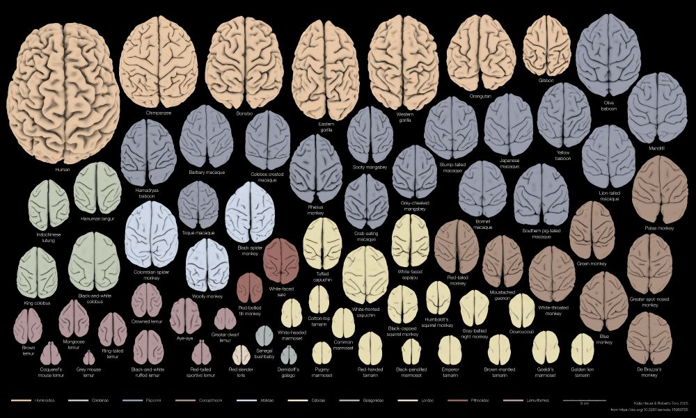
To predict the behaviour of a primate, would you rather base your guess on a closely related species or one with a similar brain shape? We looked at brains & behaviours of 70 species, you’ll be surprised!
🧵Thread on our new preprint with @r3rt0.bsky.social , doi.org/10.1101/2025...
To predict the behaviour of a primate, would you rather base your guess on a closely related species or one with a similar brain shape? We looked at brains & behaviours of 70 species, you’ll be surprised!
🧵Thread on our new preprint with @r3rt0.bsky.social , doi.org/10.1101/2025...
July 27, 2025 at 5:26 PM
1
To predict the behaviour of a primate, would you rather base your guess on a closely related species or one with a similar brain shape? We looked at brains & behaviours of 70 species, you’ll be surprised!
🧵Thread on our new preprint with @r3rt0.bsky.social , doi.org/10.1101/2025...
To predict the behaviour of a primate, would you rather base your guess on a closely related species or one with a similar brain shape? We looked at brains & behaviours of 70 species, you’ll be surprised!
🧵Thread on our new preprint with @r3rt0.bsky.social , doi.org/10.1101/2025...
Reposted by Polina Rusina
Love this!
Tooting my own horn here, but if you’re interested in some of the practical considerations mentioned at the end of the article, you can check out this perspective from earlier in my postdoc: www.cell.com/neuron/fullt...
Tooting my own horn here, but if you’re interested in some of the practical considerations mentioned at the end of the article, you can check out this perspective from earlier in my postdoc: www.cell.com/neuron/fullt...
July 18, 2025 at 5:01 PM
Love this!
Tooting my own horn here, but if you’re interested in some of the practical considerations mentioned at the end of the article, you can check out this perspective from earlier in my postdoc: www.cell.com/neuron/fullt...
Tooting my own horn here, but if you’re interested in some of the practical considerations mentioned at the end of the article, you can check out this perspective from earlier in my postdoc: www.cell.com/neuron/fullt...
Reposted by Polina Rusina
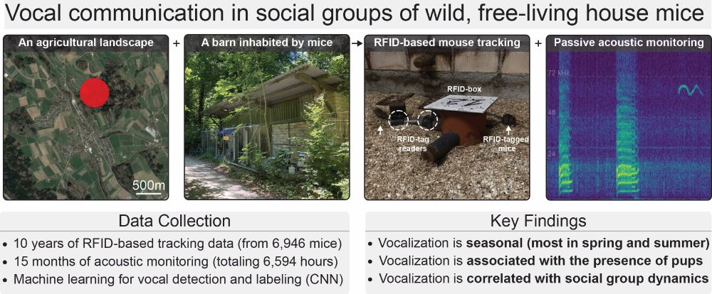
Very happy to share the latest from my postdoc!
10 yrs of mouse social networks + 1.25 yrs of acoustic data ➡️ insight into vocalization & sociality in a wild population of your favorite lab model 🐁
paper: bit.ly/4n93yyD
data: bit.ly/4lfFBEk
code: bit.ly/4kNnMwx
#bioacoustics #neuroskyence
1/8
10 yrs of mouse social networks + 1.25 yrs of acoustic data ➡️ insight into vocalization & sociality in a wild population of your favorite lab model 🐁
paper: bit.ly/4n93yyD
data: bit.ly/4lfFBEk
code: bit.ly/4kNnMwx
#bioacoustics #neuroskyence
1/8
June 18, 2025 at 6:26 PM
Very happy to share the latest from my postdoc!
10 yrs of mouse social networks + 1.25 yrs of acoustic data ➡️ insight into vocalization & sociality in a wild population of your favorite lab model 🐁
paper: bit.ly/4n93yyD
data: bit.ly/4lfFBEk
code: bit.ly/4kNnMwx
#bioacoustics #neuroskyence
1/8
10 yrs of mouse social networks + 1.25 yrs of acoustic data ➡️ insight into vocalization & sociality in a wild population of your favorite lab model 🐁
paper: bit.ly/4n93yyD
data: bit.ly/4lfFBEk
code: bit.ly/4kNnMwx
#bioacoustics #neuroskyence
1/8
Reposted by Polina Rusina
It’s a quill? A sea pen? No—it’s a gill filament! 🪶🪸🐟
Under the microscope, this fish gill structure looks just like a sea pen.
Fascinating how nature keeps circling back to the same shapes.
Want to learn how gills develop?
👉 www.biorxiv.org/content/10.1...
#FluorescenceFriday #Zebrafish
Under the microscope, this fish gill structure looks just like a sea pen.
Fascinating how nature keeps circling back to the same shapes.
Want to learn how gills develop?
👉 www.biorxiv.org/content/10.1...
#FluorescenceFriday #Zebrafish


June 20, 2025 at 1:30 PM
It’s a quill? A sea pen? No—it’s a gill filament! 🪶🪸🐟
Under the microscope, this fish gill structure looks just like a sea pen.
Fascinating how nature keeps circling back to the same shapes.
Want to learn how gills develop?
👉 www.biorxiv.org/content/10.1...
#FluorescenceFriday #Zebrafish
Under the microscope, this fish gill structure looks just like a sea pen.
Fascinating how nature keeps circling back to the same shapes.
Want to learn how gills develop?
👉 www.biorxiv.org/content/10.1...
#FluorescenceFriday #Zebrafish
Reposted by Polina Rusina

#neuroskyence folks: as my postdoc grant is running out soon, I am looking for new opportunities in systems neuroscience!
Keywords: patch clamp ephys, opto, mouse behavior, (in vivo) voltage imaging. Would love to return to the Basal Ganglia.
Sharing appreciated, and happy #FluorescenceFriday !
Keywords: patch clamp ephys, opto, mouse behavior, (in vivo) voltage imaging. Would love to return to the Basal Ganglia.
Sharing appreciated, and happy #FluorescenceFriday !
June 13, 2025 at 1:33 PM
#neuroskyence folks: as my postdoc grant is running out soon, I am looking for new opportunities in systems neuroscience!
Keywords: patch clamp ephys, opto, mouse behavior, (in vivo) voltage imaging. Would love to return to the Basal Ganglia.
Sharing appreciated, and happy #FluorescenceFriday !
Keywords: patch clamp ephys, opto, mouse behavior, (in vivo) voltage imaging. Would love to return to the Basal Ganglia.
Sharing appreciated, and happy #FluorescenceFriday !
Reposted by Polina Rusina
My lab at @ethzurich.bsky.social is looking for a motivated PhD student. We develop chemical tools for advanced fluorescence microscopy 🔬 and work at the interface of synthetic chemistry ⚗️ and protein engineering 🦠. Sharing with skilled Master students appreciated. More info at tinyurl.com/2dbjk5ty

April 8, 2025 at 8:06 AM
My lab at @ethzurich.bsky.social is looking for a motivated PhD student. We develop chemical tools for advanced fluorescence microscopy 🔬 and work at the interface of synthetic chemistry ⚗️ and protein engineering 🦠. Sharing with skilled Master students appreciated. More info at tinyurl.com/2dbjk5ty
Reposted by Polina Rusina

🚨iGluSnFR4 is finally out!🚨🧪
We present iGluSnFR4f and 4s, a novel pair of genetically-encoded glutamate indicators designed for high-fidelity imaging of synaptic activity in the living brain. ⬇️
www.biorxiv.org/content/10.1...
🎥 Below: iGluSnFR4s detecting minis in cultures w/ TTX
#Neuroscience
We present iGluSnFR4f and 4s, a novel pair of genetically-encoded glutamate indicators designed for high-fidelity imaging of synaptic activity in the living brain. ⬇️
www.biorxiv.org/content/10.1...
🎥 Below: iGluSnFR4s detecting minis in cultures w/ TTX
#Neuroscience
March 25, 2025 at 12:10 PM
🚨iGluSnFR4 is finally out!🚨🧪
We present iGluSnFR4f and 4s, a novel pair of genetically-encoded glutamate indicators designed for high-fidelity imaging of synaptic activity in the living brain. ⬇️
www.biorxiv.org/content/10.1...
🎥 Below: iGluSnFR4s detecting minis in cultures w/ TTX
#Neuroscience
We present iGluSnFR4f and 4s, a novel pair of genetically-encoded glutamate indicators designed for high-fidelity imaging of synaptic activity in the living brain. ⬇️
www.biorxiv.org/content/10.1...
🎥 Below: iGluSnFR4s detecting minis in cultures w/ TTX
#Neuroscience
Reposted by Polina Rusina
🚀 Excited to share our latest work in #JACS on eSylites!
—Synthetic, high-affinity #ChemicalBiology probes for #SuperResolution #Synapse visualization & precise mapping in neurons and brain slices—without the need for antibodies, tags, or transfection!
📢 Read more: pubs.acs.org/doi/10.1021/...
—Synthetic, high-affinity #ChemicalBiology probes for #SuperResolution #Synapse visualization & precise mapping in neurons and brain slices—without the need for antibodies, tags, or transfection!
📢 Read more: pubs.acs.org/doi/10.1021/...
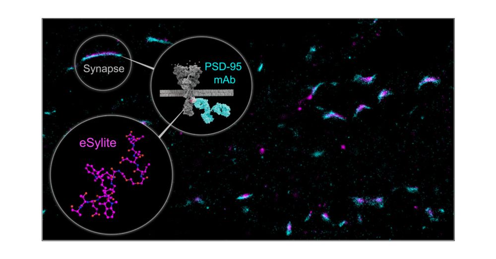
eSylites: Synthetic Probes for Visualization and Topographic Mapping of Single Excitatory Synapses
The spatiotemporal organization of the postsynaptic density (PSD) is a fundamental determinant of synaptic transmission, information processing, and storage in the brain. The major bottleneck that prevents the direct and precise representation of the nanometer-scaled organization of excitatory glutamatergic synapses is the size of antibodies, nanobodies, and the genetically encoded fluorescent tags. Here, we introduce small, high affinity synthetic probes for simplified, high contrast visualization of excitatory synapses without the limitations of larger biomolecules. In vitro binding quantification together with microscopy-based evaluation identified eSylites, a series of fluorescent bivalent peptides comprising a dye, linker, and sequence composition that show remarkable cellular target selectivity. Applied on primary neurons or brain slices at nanomolar concentrations, eSylites specifically report PSD-95, the key orchestrator of glutamate receptor nanodomains juxtaposed to the presynaptic glutamate release sites that mediate fast synaptic transmission. The eSylite design minimizes a spatial dye offset and thereby enables visualization of PSD-95 with improved localization precision and further time-resolved discrimination. In particular, we find that individual dendritic spines can contain separate nanodomains enriched for either PSD-95 or its closest homologues, PSD-93 or SAP102. Collectively, these data establish eSylites as a broadly applicable tool for simplified excitatory synapse visualization, as well as a high-end microscopy compatible probe for resolving the PSD organization with unprecedented resolution.
pubs.acs.org
March 20, 2025 at 5:21 PM
🚀 Excited to share our latest work in #JACS on eSylites!
—Synthetic, high-affinity #ChemicalBiology probes for #SuperResolution #Synapse visualization & precise mapping in neurons and brain slices—without the need for antibodies, tags, or transfection!
📢 Read more: pubs.acs.org/doi/10.1021/...
—Synthetic, high-affinity #ChemicalBiology probes for #SuperResolution #Synapse visualization & precise mapping in neurons and brain slices—without the need for antibodies, tags, or transfection!
📢 Read more: pubs.acs.org/doi/10.1021/...
Reposted by Polina Rusina
Calling all...
🔬 Microscopists
💻 BioImage Analysts
👩🔬 Life Scientists
📅 Save the Date
The 5th edition of the Crick BioImage Analysis Symposium will take place on November 24/25th 2025.
More details to follow...
#CBIAS2025
🔬 Microscopists
💻 BioImage Analysts
👩🔬 Life Scientists
📅 Save the Date
The 5th edition of the Crick BioImage Analysis Symposium will take place on November 24/25th 2025.
More details to follow...
#CBIAS2025

March 17, 2025 at 8:19 PM
Calling all...
🔬 Microscopists
💻 BioImage Analysts
👩🔬 Life Scientists
📅 Save the Date
The 5th edition of the Crick BioImage Analysis Symposium will take place on November 24/25th 2025.
More details to follow...
#CBIAS2025
🔬 Microscopists
💻 BioImage Analysts
👩🔬 Life Scientists
📅 Save the Date
The 5th edition of the Crick BioImage Analysis Symposium will take place on November 24/25th 2025.
More details to follow...
#CBIAS2025
Reposted by Polina Rusina

We must learn to showcase the practical significance of basic research, demonstrating how seemingly arcane investigations yield transformative applications, writes @tonyzador.bsky.social www.thetransmitter.org/outreach/how...
How to communicate the value of curiosity-driven research
The burden of proof is on us, as researchers, to explain why what we do is valuable to society.
www.thetransmitter.org
March 4, 2025 at 3:16 PM
We must learn to showcase the practical significance of basic research, demonstrating how seemingly arcane investigations yield transformative applications, writes @tonyzador.bsky.social www.thetransmitter.org/outreach/how...
Reposted by Polina Rusina

#RBPs and #glycoRNAs are amazing! Congrats! Great work!
#RNA #RNAsky
RNA-binding proteins and glycoRNAs form domains on the cell surface for cell-penetrating peptide entry: Cell www.cell.com/cell/fulltex...
#RNA #RNAsky
RNA-binding proteins and glycoRNAs form domains on the cell surface for cell-penetrating peptide entry: Cell www.cell.com/cell/fulltex...

RNA-binding proteins and glycoRNAs form domains on the cell surface for cell-penetrating peptide entry
Mammalian cells present RNA-binding proteins on the cell surface that form clustered
domains containing glycoRNAs.
www.cell.com
February 28, 2025 at 12:26 PM
#RBPs and #glycoRNAs are amazing! Congrats! Great work!
#RNA #RNAsky
RNA-binding proteins and glycoRNAs form domains on the cell surface for cell-penetrating peptide entry: Cell www.cell.com/cell/fulltex...
#RNA #RNAsky
RNA-binding proteins and glycoRNAs form domains on the cell surface for cell-penetrating peptide entry: Cell www.cell.com/cell/fulltex...
Reposted by Polina Rusina

nature.com/articles/s41467-025-57053-9
Finally this work is out - we show that short sequence motifs direct proteins to specific peroxisomal sub-domains. Fantastic collaboration with @bangebalcony.bsky.social , Johannes Freitag @unimarburg.bsky.social and many more! @uio.no @cancelluio.bsky.social
Finally this work is out - we show that short sequence motifs direct proteins to specific peroxisomal sub-domains. Fantastic collaboration with @bangebalcony.bsky.social , Johannes Freitag @unimarburg.bsky.social and many more! @uio.no @cancelluio.bsky.social

Peroxisomal core structures segregate diverse metabolic pathways - Nature Communications
Peroxisomes contain detergent resistant core structures. Bäcker et al. show that these core structures contain diverse enzymes and serve to enable metabolic compartmentalization of the peroxisome lume...
nature.com
February 20, 2025 at 5:02 PM
nature.com/articles/s41467-025-57053-9
Finally this work is out - we show that short sequence motifs direct proteins to specific peroxisomal sub-domains. Fantastic collaboration with @bangebalcony.bsky.social , Johannes Freitag @unimarburg.bsky.social and many more! @uio.no @cancelluio.bsky.social
Finally this work is out - we show that short sequence motifs direct proteins to specific peroxisomal sub-domains. Fantastic collaboration with @bangebalcony.bsky.social , Johannes Freitag @unimarburg.bsky.social and many more! @uio.no @cancelluio.bsky.social
Reposted by Polina Rusina


Reposted by Polina Rusina

Making intrabodies from antibodies just got easier! Learn how we made 𝟭𝟵 intrabodies to bind and light up peptides and histone modifications in live cells. And thanks to Academia, all sequences are freely available. (video credit: Yuko Sato @YukoSatoT2) (1/15)
www.biorxiv.org/content/10.1...
www.biorxiv.org/content/10.1...
February 12, 2025 at 6:28 AM
Making intrabodies from antibodies just got easier! Learn how we made 𝟭𝟵 intrabodies to bind and light up peptides and histone modifications in live cells. And thanks to Academia, all sequences are freely available. (video credit: Yuko Sato @YukoSatoT2) (1/15)
www.biorxiv.org/content/10.1...
www.biorxiv.org/content/10.1...
Reposted by Polina Rusina
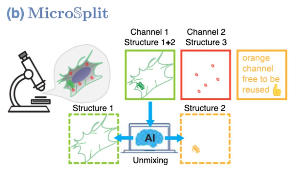
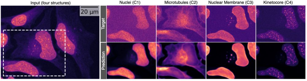
Immagine you could image two cellular structures in the same fluorescent channel and still reliably get them separated afterwards…
What would you do with this?
Now… what would you do if that also worked with 4 structures at once? 👇 #MicroSplit #preview🧵
What would you do with this?
Now… what would you do if that also worked with 4 structures at once? 👇 #MicroSplit #preview🧵
February 8, 2025 at 7:49 AM
Immagine you could image two cellular structures in the same fluorescent channel and still reliably get them separated afterwards…
What would you do with this?
Now… what would you do if that also worked with 4 structures at once? 👇 #MicroSplit #preview🧵
What would you do with this?
Now… what would you do if that also worked with 4 structures at once? 👇 #MicroSplit #preview🧵
Reposted by Polina Rusina
Protein codes promote selective subcellular compartmentalization | Science www.science.org/doi/10.1126/...

Protein codes promote selective subcellular compartmentalization
Cells have evolved mechanisms to distribute ~10 billion protein molecules to subcellular compartments where diverse proteins involved in shared functions must assemble. Here, we demonstrate that proteins with shared functions share amino acid sequence ...
www.science.org
February 6, 2025 at 7:17 PM
Protein codes promote selective subcellular compartmentalization | Science www.science.org/doi/10.1126/...
Reposted by Polina Rusina
Answering the call of @haesleinhuepf.bsky.social, Christian Tischer, Pete Bankhead, @kbias.bsky.social @bethcimini.bsky.social
My Fiji training notes are now FAIR. All online in an open format to download and use for teaching and training. All in Google Doc or PDF formats
bit.ly/4hkfEBF
My Fiji training notes are now FAIR. All online in an open format to download and use for teaching and training. All in Google Doc or PDF formats
bit.ly/4hkfEBF

Home
This site contains the complete version of the Fiji Training Manual that is written and maintained by Cameron Nowell of the Monash Institue of Pharmaceutical Science.
This manual is provided under the...
bit.ly
February 5, 2025 at 10:57 PM
Answering the call of @haesleinhuepf.bsky.social, Christian Tischer, Pete Bankhead, @kbias.bsky.social @bethcimini.bsky.social
My Fiji training notes are now FAIR. All online in an open format to download and use for teaching and training. All in Google Doc or PDF formats
bit.ly/4hkfEBF
My Fiji training notes are now FAIR. All online in an open format to download and use for teaching and training. All in Google Doc or PDF formats
bit.ly/4hkfEBF
Reposted by Polina Rusina

Do you know Daisy Roulland-Dussoix? She is one of the discoverers of restriction enzymes, who’s findings paved the way for the development of recombinant DNA and cloning technologies. Accordingly, the finding was rewarded with a #NobelPrize. But the prize didn’t go to her.
🧵👇
🧵👇

February 1, 2025 at 3:29 PM
Do you know Daisy Roulland-Dussoix? She is one of the discoverers of restriction enzymes, who’s findings paved the way for the development of recombinant DNA and cloning technologies. Accordingly, the finding was rewarded with a #NobelPrize. But the prize didn’t go to her.
🧵👇
🧵👇
Reposted by Polina Rusina

A pyramidal neuron from the prefrontal cortex. #microscopymonday
Labelled with DiI, imaged with the FV3000 CLSM, & depicted using @kwolbachia.bsky.social new KTZ_bw_kawa LUT.
Labelled with DiI, imaged with the FV3000 CLSM, & depicted using @kwolbachia.bsky.social new KTZ_bw_kawa LUT.

January 27, 2025 at 5:38 AM
A pyramidal neuron from the prefrontal cortex. #microscopymonday
Labelled with DiI, imaged with the FV3000 CLSM, & depicted using @kwolbachia.bsky.social new KTZ_bw_kawa LUT.
Labelled with DiI, imaged with the FV3000 CLSM, & depicted using @kwolbachia.bsky.social new KTZ_bw_kawa LUT.
Reposted by Polina Rusina
There are new stable red fluorescent proteins coming, but organic fluorophores are fighting back! Impressive photostability of Phoenix Fluor 555 for live-cell imaging with HaloTag, just out in @naturemethods.bsky.social:
doi.org/10.1038/s415...
doi.org/10.1038/s415...
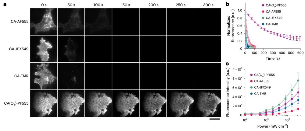
January 16, 2025 at 10:52 AM
There are new stable red fluorescent proteins coming, but organic fluorophores are fighting back! Impressive photostability of Phoenix Fluor 555 for live-cell imaging with HaloTag, just out in @naturemethods.bsky.social:
doi.org/10.1038/s415...
doi.org/10.1038/s415...