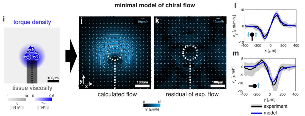
We quantified these chiral tissue flows in 25 quail embryos and inferred forces using a fluid model, suggesting a torque of 6μNμm is generated locally at the node.
We quantified these chiral tissue flows in 25 quail embryos and inferred forces using a fluid model, suggesting a torque of 6μNμm is generated locally at the node.
We quantified these chiral tissue flows in 25 quail embryos and inferred forces using a fluid model, suggesting a torque of 6μNμm is generated locally at the node.
Jerome found cells moving to the left around the node, repositioning a Shh domain to the left, but what makes them do that?

Jerome found cells moving to the left around the node, repositioning a Shh domain to the left, but what makes them do that?

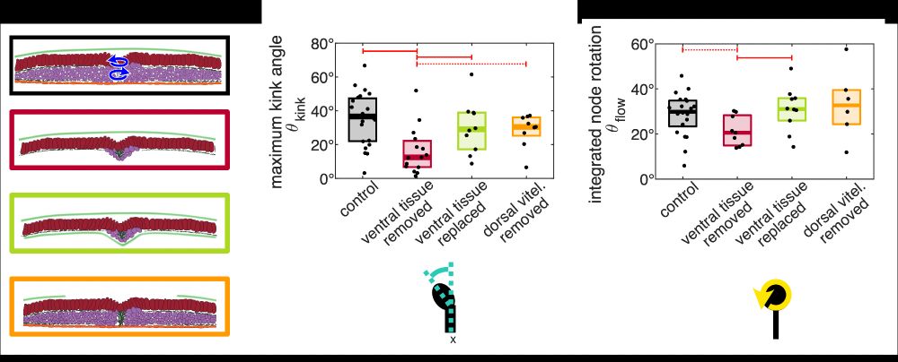
But now comes the really striking result: She found that replacing the meso-/endo tissue with a vitel. membrane is sufficient to restore node rotation. So, you simply need some mech. substrate. sustaining a counter-torque such that the node can drive its own rotation.

But now comes the really striking result: She found that replacing the meso-/endo tissue with a vitel. membrane is sufficient to restore node rotation. So, you simply need some mech. substrate. sustaining a counter-torque such that the node can drive its own rotation.
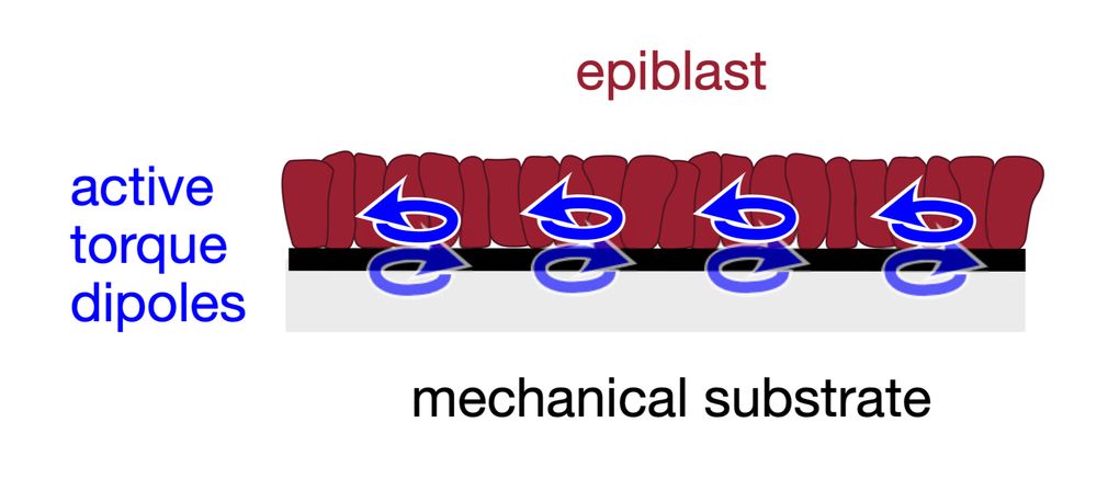
So, the following picture following mechanical picture of avian L/R sym. break. emerges: actomyosin-activity generates cell-scale torque dipoles that propel the rotation of node and surrounding epiblast with respect to some rigid substrate.


So, the following picture following mechanical picture of avian L/R sym. break. emerges: actomyosin-activity generates cell-scale torque dipoles that propel the rotation of node and surrounding epiblast with respect to some rigid substrate.
But is the node really capable of making itself rotate? Lasercuts by Julia show yes, providing maybe the first direct evidence for a tissue-scale active chiral torque. Also, we show that the apparent torque is actomyosin-dependent, consistent with previous observations by Jerome.

But is the node really capable of making itself rotate? Lasercuts by Julia show yes, providing maybe the first direct evidence for a tissue-scale active chiral torque. Also, we show that the apparent torque is actomyosin-dependent, consistent with previous observations by Jerome.
We quantified these chiral tissue flows in 25 quail embryos and inferred forces using a fluid model, suggesting a torque of 6μNμm is generated locally at the node.
We quantified these chiral tissue flows in 25 quail embryos and inferred forces using a fluid model, suggesting a torque of 6μNμm is generated locally at the node.
Why is the heart on the left? Somehow, embryos mange to translate mol. chirality to tissue-scale handedness. In various vertebrates, that’s the job of cilia. But birds, chameleons, pigs do it differently, as shown before by the fabulous @jeromegros.bsky.social and @shylonatasha.bsky.social
Why is the heart on the left? Somehow, embryos mange to translate mol. chirality to tissue-scale handedness. In various vertebrates, that’s the job of cilia. But birds, chameleons, pigs do it differently, as shown before by the fabulous @jeromegros.bsky.social and @shylonatasha.bsky.social

