


Michalis Averof
@michalis-averof.bsky.social
Comparative developmental biology, regeneration, non-conventional model organisms, live imaging; see www.averof-lab.org
That's what 'the napolitans' we up to when they briefly invaded our lab! Wonderful memories from December 2021 and November 2022. Congratulations!!


November 5, 2025 at 8:11 PM
That's what 'the napolitans' we up to when they briefly invaded our lab! Wonderful memories from December 2021 and November 2022. Congratulations!!
Our ultimate goal: exploring biology beyond well established research organisms, by building tools that will make new things visible
Ending 🧵 with a marker for visualising chanoflagellates, closest unicellular relatives of animals, by @jujumathieu.bsky.social and @thibautbrunet.bsky.social
Ending 🧵 with a marker for visualising chanoflagellates, closest unicellular relatives of animals, by @jujumathieu.bsky.social and @thibautbrunet.bsky.social

October 27, 2025 at 11:19 PM
Our ultimate goal: exploring biology beyond well established research organisms, by building tools that will make new things visible
Ending 🧵 with a marker for visualising chanoflagellates, closest unicellular relatives of animals, by @jujumathieu.bsky.social and @thibautbrunet.bsky.social
Ending 🧵 with a marker for visualising chanoflagellates, closest unicellular relatives of animals, by @jujumathieu.bsky.social and @thibautbrunet.bsky.social
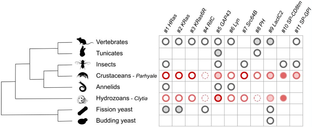
By studying these markers in a multiple organisms, we could identify ones that label the cell membrane most clearly and consistently across species (circle sizes represent how well markers perform in each species). Gathering the info involved more than 10 research labs.
see doi.org/10.1101/2024... 🧵
see doi.org/10.1101/2024... 🧵

October 27, 2025 at 10:05 PM
By studying these markers in a multiple organisms, we could identify ones that label the cell membrane most clearly and consistently across species (circle sizes represent how well markers perform in each species). Gathering the info involved more than 10 research labs.
see doi.org/10.1101/2024... 🧵
see doi.org/10.1101/2024... 🧵
Some of the markers do not label the cell surface clearly, but shuttle across different parts of the cell. You can see in this early crustacean embryo, how the fluorescence pattern changes as the cells multiply. The video was made by Manon Koenig, Irene Karapidaki and @berylbiologist.bsky.social
🧵
🧵
October 27, 2025 at 9:36 PM
Some of the markers do not label the cell surface clearly, but shuttle across different parts of the cell. You can see in this early crustacean embryo, how the fluorescence pattern changes as the cells multiply. The video was made by Manon Koenig, Irene Karapidaki and @berylbiologist.bsky.social
🧵
🧵
Our lab studies how animals regenerate their body, e.g. how crustaceans regenerate broken legs. One of our aims is to understand if regeneration re-uses the gene networks that built the legs in the first place. Arthur Monternier, an artist in our team, captured the question in this cartoon.

October 27, 2025 at 3:25 PM
Our lab studies how animals regenerate their body, e.g. how crustaceans regenerate broken legs. One of our aims is to understand if regeneration re-uses the gene networks that built the legs in the first place. Arthur Monternier, an artist in our team, captured the question in this cartoon.
And this is the surface of the larvae of mediterranean stinging jellyfish. It's composed of cells measuring about a hundredth of a millimetre in size. The cells' outlines are visible thanks to fluorescent markers identified by Clara Deleau and @cnidevo.bsky.social
see doi.org/10.1101/2024...
🧵
see doi.org/10.1101/2024...
🧵

October 27, 2025 at 11:52 AM
And this is the surface of the larvae of mediterranean stinging jellyfish. It's composed of cells measuring about a hundredth of a millimetre in size. The cells' outlines are visible thanks to fluorescent markers identified by Clara Deleau and @cnidevo.bsky.social
see doi.org/10.1101/2024...
🧵
see doi.org/10.1101/2024...
🧵
These are embryos of a jellyfish, made up of cells that are about a fiftieth of a millimetre in size. Their outlines are visible thanks to fluorescent markers identified by Sarah Asaf and @clytia-vlfr.bsky.social
see doi.org/10.1101/2024...
🧵
see doi.org/10.1101/2024...
🧵

October 26, 2025 at 2:12 PM
These are embryos of a jellyfish, made up of cells that are about a fiftieth of a millimetre in size. Their outlines are visible thanks to fluorescent markers identified by Sarah Asaf and @clytia-vlfr.bsky.social
see doi.org/10.1101/2024...
🧵
see doi.org/10.1101/2024...
🧵
What are these?
You are looking at embryos of a sea squirt. Each of the 'soap bubbles' is a living cell, about a fourtieth of a millimetre in size. The outlines of the cells are visible thanks to fluorescent markers identified by Hitoyoshi Yasuo @hitoyas.bsky.social
see doi.org/10.1101/2024... 🧵
You are looking at embryos of a sea squirt. Each of the 'soap bubbles' is a living cell, about a fourtieth of a millimetre in size. The outlines of the cells are visible thanks to fluorescent markers identified by Hitoyoshi Yasuo @hitoyas.bsky.social
see doi.org/10.1101/2024... 🧵

October 26, 2025 at 8:55 AM
What are these?
You are looking at embryos of a sea squirt. Each of the 'soap bubbles' is a living cell, about a fourtieth of a millimetre in size. The outlines of the cells are visible thanks to fluorescent markers identified by Hitoyoshi Yasuo @hitoyas.bsky.social
see doi.org/10.1101/2024... 🧵
You are looking at embryos of a sea squirt. Each of the 'soap bubbles' is a living cell, about a fourtieth of a millimetre in size. The outlines of the cells are visible thanks to fluorescent markers identified by Hitoyoshi Yasuo @hitoyas.bsky.social
see doi.org/10.1101/2024... 🧵
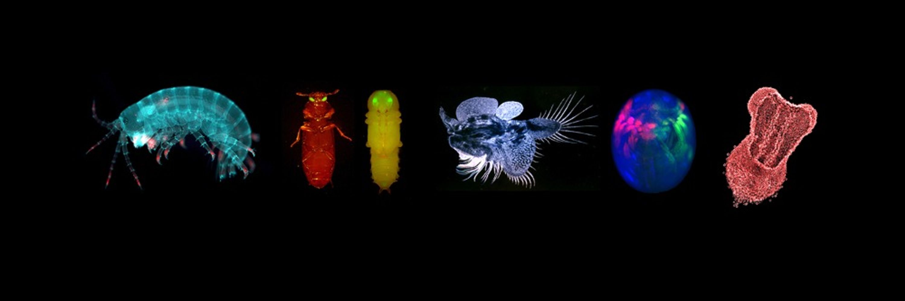
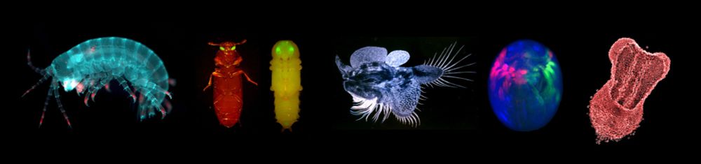
Project started a year ago, when we gathered a set of 11 tags, made them easy to test as mRNA that can be injected in eggs, and sent to 30 labs around the world. We present the results of testing these tags in animals as diverse as sea urchin, beetle, crustacean, worm, sea anemone & jellyfish.
2/4
2/4
October 23, 2025 at 8:16 AM
Project started a year ago, when we gathered a set of 11 tags, made them easy to test as mRNA that can be injected in eggs, and sent to 30 labs around the world. We present the results of testing these tags in animals as diverse as sea urchin, beetle, crustacean, worm, sea anemone & jellyfish.
2/4
2/4
X-ray imaging of crustacean legs at the DESY synchrotron, Hamburg. Cross-section of regenerating Parhyale leg (~1/10 of a mm in width). X-rays give accurate 3D view with almost cellular resolution. With Angelika Svetlove, Kevin Grüner & Mathilde Paris @zoocell.biologists.social.ap.brid.gy


September 24, 2025 at 1:17 PM
X-ray imaging of crustacean legs at the DESY synchrotron, Hamburg. Cross-section of regenerating Parhyale leg (~1/10 of a mm in width). X-rays give accurate 3D view with almost cellular resolution. With Angelika Svetlove, Kevin Grüner & Mathilde Paris @zoocell.biologists.social.ap.brid.gy
Interested in a sabbatical in Lyon? The Collegium de Lyon provides accommodation and attractive interdisciplinary environment for 1 or 2 semesters. Open to all disciplines. You can be affiliated with any local department/lab. Apply now for 2026-27. collegium.universite-lyon.fr/2026-2027-un...

September 2, 2025 at 8:45 AM
Interested in a sabbatical in Lyon? The Collegium de Lyon provides accommodation and attractive interdisciplinary environment for 1 or 2 semesters. Open to all disciplines. You can be affiliated with any local department/lab. Apply now for 2026-27. collegium.universite-lyon.fr/2026-2027-un...
Latest paper elifesciences.org/articles/107... closes an important cycle in our efforts to study regeneration: week-long recordings allow us to observe the behaviour of cells during the entire course of regeneration in a crustacean leg – bright objects in movie are fluorescent nuclei of cells. 1/6
August 8, 2025 at 5:39 PM
Latest paper elifesciences.org/articles/107... closes an important cycle in our efforts to study regeneration: week-long recordings allow us to observe the behaviour of cells during the entire course of regeneration in a crustacean leg – bright objects in movie are fluorescent nuclei of cells. 1/6
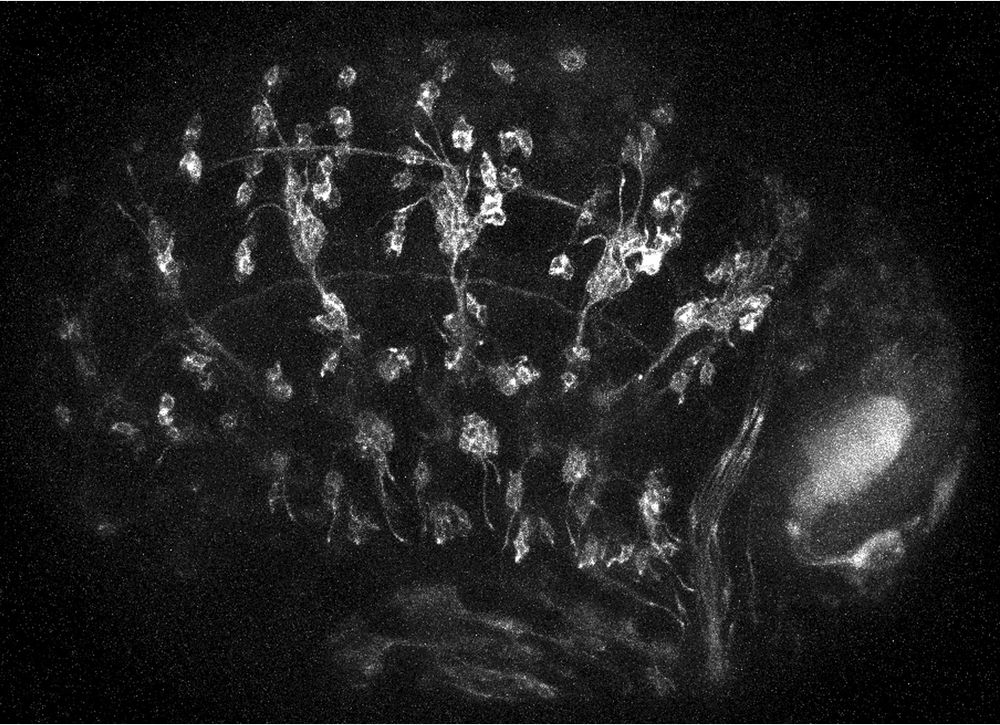
for light sheet microscopy w. Flamingo team @metteh-thorsager.bsky.social, genetic barcoding w. @mgrillo.bsky.social, and a video game for cell tracking w. @jytinevez.bsky.social. We'll apply these tools to discover the progenitors for sense organ regeneration in Parhyale. 2/3
July 13, 2025 at 2:55 PM
for light sheet microscopy w. Flamingo team @metteh-thorsager.bsky.social, genetic barcoding w. @mgrillo.bsky.social, and a video game for cell tracking w. @jytinevez.bsky.social. We'll apply these tools to discover the progenitors for sense organ regeneration in Parhyale. 2/3
We've just been awarded a grant to study the cellular basis of regeneration – to track the progenitors of sensory organs in the context of leg regeneration, in our favourite crustacean tinyurl.com/parhyale, based on live imaging and cell tracking. The project involves some cool collaborations... 1/3
July 13, 2025 at 2:55 PM
We've just been awarded a grant to study the cellular basis of regeneration – to track the progenitors of sensory organs in the context of leg regeneration, in our favourite crustacean tinyurl.com/parhyale, based on live imaging and cell tracking. The project involves some cool collaborations... 1/3
diverse body shapes of Trichoplax, from small pancake to irregular spaghetti
June 25, 2025 at 2:50 PM
diverse body shapes of Trichoplax, from small pancake to irregular spaghetti
Trichoplax adhaerens is one of the simplest and most enigmatic animals on earth. Its body shows incredible fluidity, changing shape within minutes. In a recent visit by Andrea Pasini we had the chance to host and observe these amazing animals live.
[movie accelerated 3x; animal 0.5 to 1 mm in size]
[movie accelerated 3x; animal 0.5 to 1 mm in size]
May 10, 2025 at 5:53 PM
Trichoplax adhaerens is one of the simplest and most enigmatic animals on earth. Its body shows incredible fluidity, changing shape within minutes. In a recent visit by Andrea Pasini we had the chance to host and observe these amazing animals live.
[movie accelerated 3x; animal 0.5 to 1 mm in size]
[movie accelerated 3x; animal 0.5 to 1 mm in size]
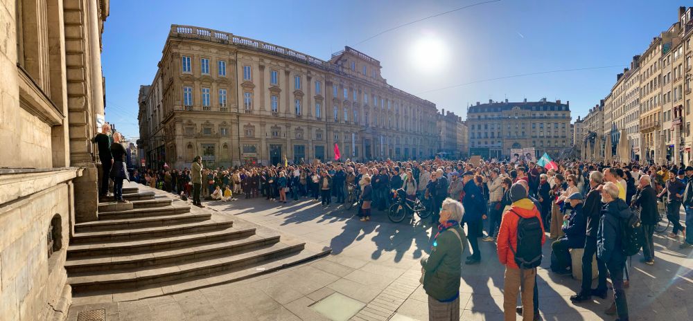
I prepared these as banners for the #StandUpForScience demonstration on Friday. Happy to share the high-res files, if anyone wants to use them.


March 5, 2025 at 7:09 PM
I prepared these as banners for the #StandUpForScience demonstration on Friday. Happy to share the high-res files, if anyone wants to use them.
Do you have experience in generating transgenic tools and fluorescent markers? Are you interested in developing new tools in a non-conventional model, to observe and genetically manipulate cells during leg regeneration? We are recruiting!
www.averof-lab.org/pages/3560
www.averof-lab.org/pages/3560
February 28, 2025 at 6:12 PM
Do you have experience in generating transgenic tools and fluorescent markers? Are you interested in developing new tools in a non-conventional model, to observe and genetically manipulate cells during leg regeneration? We are recruiting!
www.averof-lab.org/pages/3560
www.averof-lab.org/pages/3560
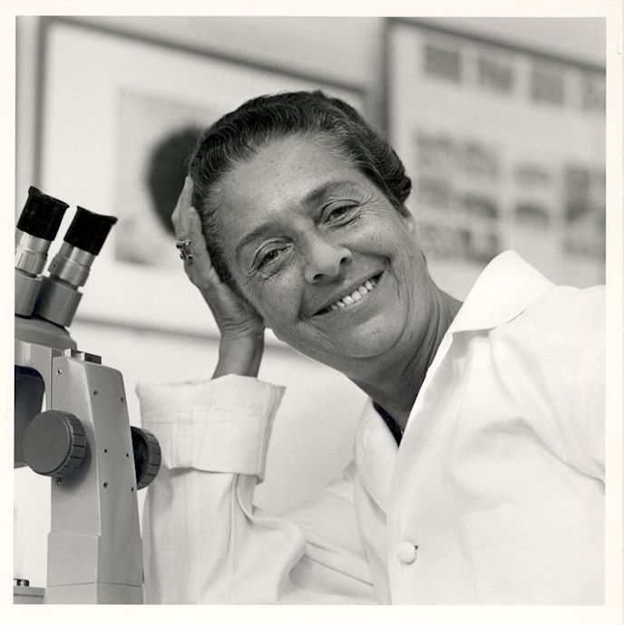
Rita Levi-Montalcini defied fascism, which excluded her from university due to her jewish ancestry, by setting up a clandestine lab in her own bedroom. During 1940-42 she carried out experiments that set the foundations for her Nobel-prize-winning research a decade later.
nautil.us/a-lab-of-her...
nautil.us/a-lab-of-her...
February 27, 2025 at 8:58 AM
Rita Levi-Montalcini defied fascism, which excluded her from university due to her jewish ancestry, by setting up a clandestine lab in her own bedroom. During 1940-42 she carried out experiments that set the foundations for her Nobel-prize-winning research a decade later.
nautil.us/a-lab-of-her...
nautil.us/a-lab-of-her...
As the world of science wakes up to modern fascism, it's worth remembering older generations who stood up for humanity in much harsher circumstances. Jacques Monod worked actively for the resistance in WWII, while pursuing early stages of his research in occupied Paris. @standupforscifr.bsky.social

February 26, 2025 at 5:40 PM
As the world of science wakes up to modern fascism, it's worth remembering older generations who stood up for humanity in much harsher circumstances. Jacques Monod worked actively for the resistance in WWII, while pursuing early stages of his research in occupied Paris. @standupforscifr.bsky.social
Would you like to join us in screening these tags in diverse species? Please get in touch. We're happy to share the constructs for generating mRNA, and will be happy to include new species –and new co-authors– in our comparative study. 3/4
November 12, 2024 at 11:05 PM
Would you like to join us in screening these tags in diverse species? Please get in touch. We're happy to share the constructs for generating mRNA, and will be happy to include new species –and new co-authors– in our comparative study. 3/4
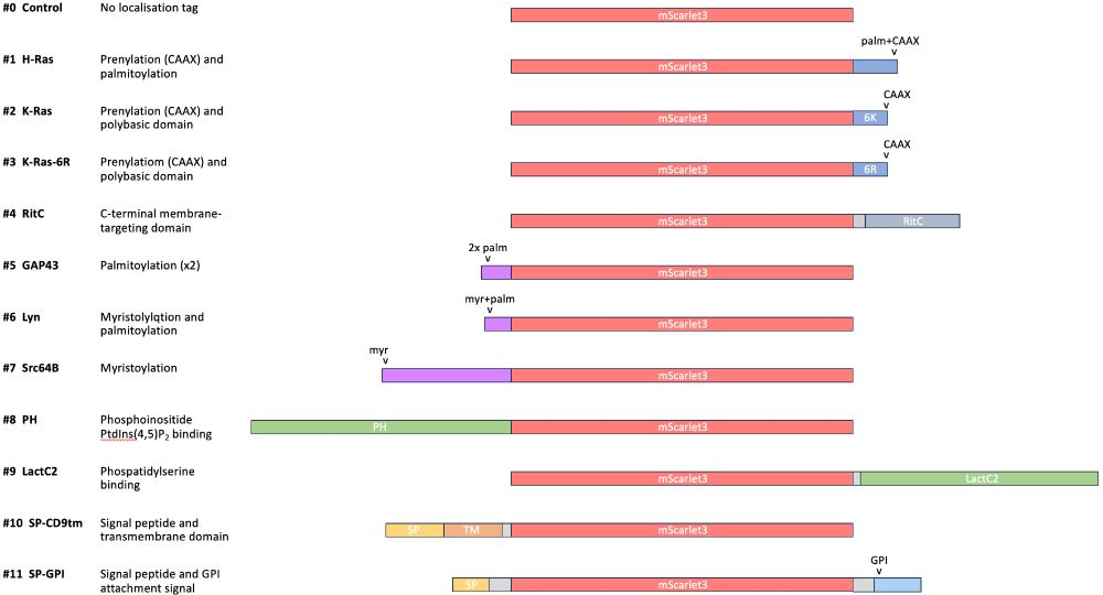
The tags employ different mechanisms for plasma membrane localisation, including prenylation, myristoylation, palmitoylation, GPI anchoring, lipid-binding domains, signal peptides and transmembrane domains for trafficking to the plasma membrane. All fused to mScarlet3 FP. 2/4
November 12, 2024 at 11:05 PM
The tags employ different mechanisms for plasma membrane localisation, including prenylation, myristoylation, palmitoylation, GPI anchoring, lipid-binding domains, signal peptides and transmembrane domains for trafficking to the plasma membrane. All fused to mScarlet3 FP. 2/4
Together with the preprint, we are releasing 22 image datasets capturing the full course of leg regeneration. This is foundational work for our research on regeneration, supported by the European Union ERC and France ANR. A multi-year collaboration with a great team! 3/3
zenodo.org/records/1370...
zenodo.org/records/1370...

September 12, 2024 at 2:55 PM
Together with the preprint, we are releasing 22 image datasets capturing the full course of leg regeneration. This is foundational work for our research on regeneration, supported by the European Union ERC and France ANR. A multi-year collaboration with a great team! 3/3
zenodo.org/records/1370...
zenodo.org/records/1370...
Looking for a membrane localisation tag? unclear what will work in your spp? We gathered 10 tags (diff mechanisms of memb localisation) + fused them with red FP. Interested in joining us for a comparative screen? We'll share plasmids for making mRNA, you can inject in your favourite model. #evodevo

July 13, 2024 at 7:18 PM
Looking for a membrane localisation tag? unclear what will work in your spp? We gathered 10 tags (diff mechanisms of memb localisation) + fused them with red FP. Interested in joining us for a comparative screen? We'll share plasmids for making mRNA, you can inject in your favourite model. #evodevo