


Kevin Staras
@kevinstaras.bsky.social
NeuroProf at University of Sussex, UK • Synapses, Circuits, Plasticity, Disease, Decision-making
https://www.thestaraslab.org/
https://www.thestaraslab.org/
Reposted by Kevin Staras

Sussex Neuroscience 4-year PhD programme www.sussex.ac.uk/study/phd/de...
Sussex Neuroscience 4-Year Programme PhD at the University of Sussex
Join a team of world-leading experts in our high-spec Neuroscience Research Centre whose aim is to understand the nervous system and various neurological disorders.
www.sussex.ac.uk
June 17, 2025 at 7:58 PM
Sussex Neuroscience 4-year PhD programme www.sussex.ac.uk/study/phd/de...
Reposted by Kevin Staras

Oops the original post was deleted! The news is here: bsky.app/profile/cham...
👏 Congratulations to @megancarey.bsky.social, principal investigator at @champalimaudr.bsky.social on winning a 2.5M€ #AdvancedGrantInLifeSciences, awarded by the @erc.europa.eu, to develop the project 🧠 SCUPTABELLUM - Sculpting cerebellar activity across timescales.

June 17, 2025 at 7:02 PM
Oops the original post was deleted! The news is here: bsky.app/profile/cham...
And here's the brain atlas link again, hopefully working this time: sites.google.com/view/snailbr...

SnailBrainMap
About The Project
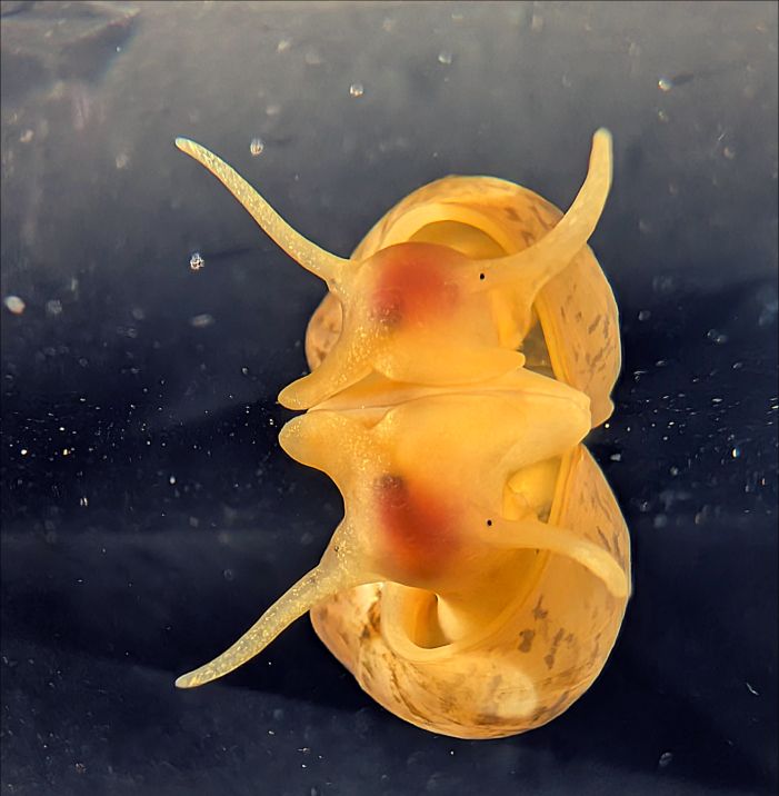
Thanks to their accessible nervous systems, molluscs have provided some of the most fundamental insights into how neural circuits generate and control behavior. Notably, their neuron...
sites.google.com
February 28, 2025 at 10:08 AM
And here's the brain atlas link again, hopefully working this time: sites.google.com/view/snailbr...
Michael Crossley led the experimental work, supported by Anna Simon, @arndroth.bsky.social and
@enzomarra.bsky.social Thanks to @sussexneuro.bsky.social @leverhulme.bsky.social @ukri.org and @diamondlightsource.bsky.social for funding support. Thanks for reading! 10/10
@enzomarra.bsky.social Thanks to @sussexneuro.bsky.social @leverhulme.bsky.social @ukri.org and @diamondlightsource.bsky.social for funding support. Thanks for reading! 10/10
February 28, 2025 at 9:40 AM
Michael Crossley led the experimental work, supported by Anna Simon, @arndroth.bsky.social and
@enzomarra.bsky.social Thanks to @sussexneuro.bsky.social @leverhulme.bsky.social @ukri.org and @diamondlightsource.bsky.social for funding support. Thanks for reading! 10/10
@enzomarra.bsky.social Thanks to @sussexneuro.bsky.social @leverhulme.bsky.social @ukri.org and @diamondlightsource.bsky.social for funding support. Thanks for reading! 10/10
Our approach should readily generalize to other model systems with comparable brain sizes (e.g. other molluscs, crustacea, annelids, insects). On its own, it won’t yield a full wiring diagram, but it does rapidly provide a detailed overview map for atlas building and comparative studies. 9/10
February 28, 2025 at 9:38 AM
Our approach should readily generalize to other model systems with comparable brain sizes (e.g. other molluscs, crustacea, annelids, insects). On its own, it won’t yield a full wiring diagram, but it does rapidly provide a detailed overview map for atlas building and comparative studies. 9/10
This provides the locations of principal feeding-circuit cell types, including motoneurons, CPG neurons and modulatory cells, alongside a detailed summary of their main functional properties. 8/10

February 28, 2025 at 9:37 AM
This provides the locations of principal feeding-circuit cell types, including motoneurons, CPG neurons and modulatory cells, alongside a detailed summary of their main functional properties. 8/10
We also brought together the anatomical mapping and functional information to establish the beginnings of a fully scalable functional cell atlas of the brain of Lymnaea stagnalis: sites.google.com/view/snailbr... 7/10
February 28, 2025 at 9:36 AM
We also brought together the anatomical mapping and functional information to establish the beginnings of a fully scalable functional cell atlas of the brain of Lymnaea stagnalis: sites.google.com/view/snailbr... 7/10
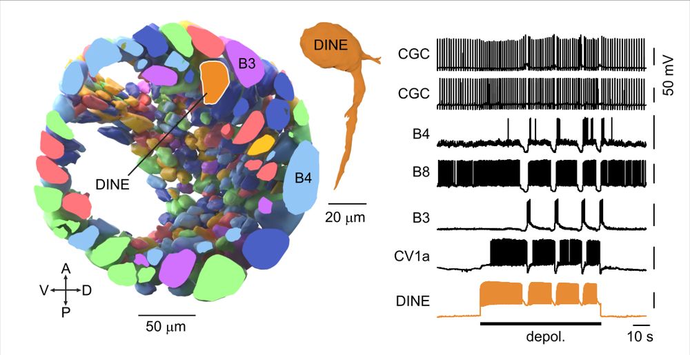
The consistent positioning of neurons across Lymnaea brains means the atlas can guide follow-up functional experiments. Targeting a non-superficial region led to the discovery of DINE (“Diamond Neuron”), an apt name 😜 because it activates the food ingestion circuitry. 6/10
February 28, 2025 at 9:35 AM
The consistent positioning of neurons across Lymnaea brains means the atlas can guide follow-up functional experiments. Targeting a non-superficial region led to the discovery of DINE (“Diamond Neuron”), an apt name 😜 because it activates the food ingestion circuitry. 6/10
The 3D reconstruction revealed the organization of neurons beneath the surface layer for the first time. It turns out around half the neurons (coloured orange) are non-superficial - a hidden world of circuit components that can now be studied. 5/10
February 28, 2025 at 9:35 AM
The 3D reconstruction revealed the organization of neurons beneath the surface layer for the first time. It turns out around half the neurons (coloured orange) are non-superficial - a hidden world of circuit components that can now be studied. 5/10
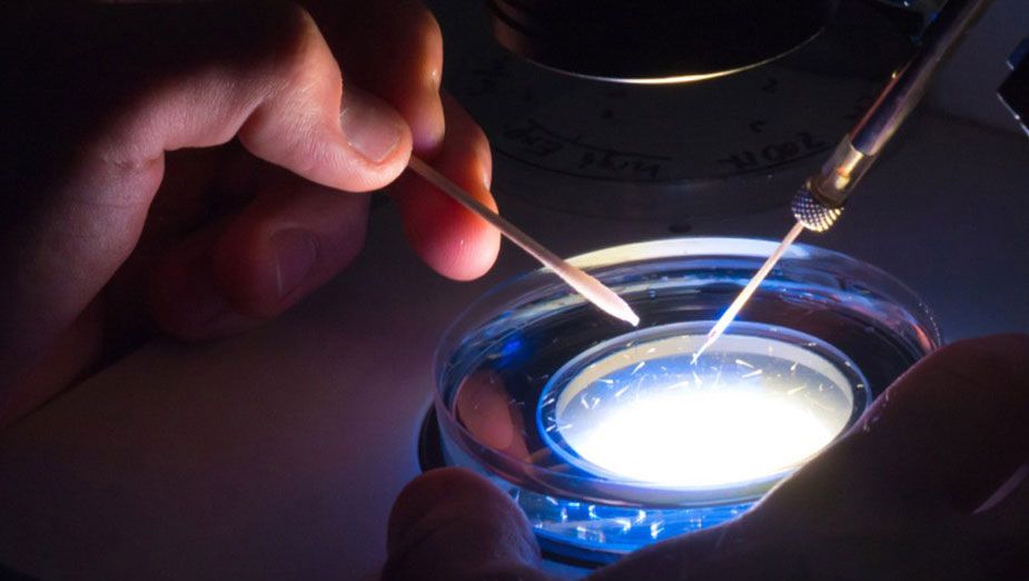
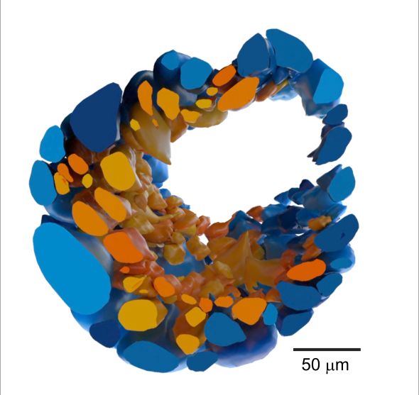
We then used the excellent volume image-sharing, annotation, and reconstruction platform
@webknossos.org to fully reconstruct the buccal ganglia (one side is shown here) housing the main feeding circuitry, yielding the first accurate estimate of the total number of neurons: ~1100. 4/10
@webknossos.org to fully reconstruct the buccal ganglia (one side is shown here) housing the main feeding circuitry, yielding the first accurate estimate of the total number of neurons: ~1100. 4/10
February 28, 2025 at 9:34 AM
We then used the excellent volume image-sharing, annotation, and reconstruction platform
@webknossos.org to fully reconstruct the buccal ganglia (one side is shown here) housing the main feeding circuitry, yielding the first accurate estimate of the total number of neurons: ~1100. 4/10
@webknossos.org to fully reconstruct the buccal ganglia (one side is shown here) housing the main feeding circuitry, yielding the first accurate estimate of the total number of neurons: ~1100. 4/10
Michael Crossley led the experimental work, supported by Anna Simon, @arndroth.bsky.social and @enzomarra.bsky.social Thanks to @sussexneuro.bsky.social @leverhulme.bsky.social @ukri.org and @diamondlightsource.bsky.social for funding support. Thanks for reading! 10/10
February 28, 2025 at 8:49 AM
Michael Crossley led the experimental work, supported by Anna Simon, @arndroth.bsky.social and @enzomarra.bsky.social Thanks to @sussexneuro.bsky.social @leverhulme.bsky.social @ukri.org and @diamondlightsource.bsky.social for funding support. Thanks for reading! 10/10
Our approach should readily generalize to other model systems with comparable brain sizes (e.g. other molluscs, crustacea, annelids, insects). On its own, it won’t yield a full wiring diagram, but it does rapidly provide a detailed overview map for atlas building and comparative studies. 9/10
February 28, 2025 at 8:47 AM
Our approach should readily generalize to other model systems with comparable brain sizes (e.g. other molluscs, crustacea, annelids, insects). On its own, it won’t yield a full wiring diagram, but it does rapidly provide a detailed overview map for atlas building and comparative studies. 9/10
This provides the locations of principal feeding-circuit cell types, including motoneurons, CPG neurons and modulatory cells, alongside a detailed summary of their main functional properties. 8/10
February 28, 2025 at 8:45 AM
This provides the locations of principal feeding-circuit cell types, including motoneurons, CPG neurons and modulatory cells, alongside a detailed summary of their main functional properties. 8/10
We also brought together the anatomical mapping and functional information to establish the beginnings of a fully scalable functional cell atlas of the brain of Lymnaea stagnalis: sites.google.com/view/snailbr... 7/10
February 28, 2025 at 8:44 AM
We also brought together the anatomical mapping and functional information to establish the beginnings of a fully scalable functional cell atlas of the brain of Lymnaea stagnalis: sites.google.com/view/snailbr... 7/10
The consistent positioning of neurons across Lymnaea brains means the atlas can guide follow-up functional experiments. Targeting a non-superficial region led to the discovery of DINE (“Diamond Neuron”), an apt name 😜 because it activates the food ingestion circuitry. 6/10
February 28, 2025 at 8:44 AM
The consistent positioning of neurons across Lymnaea brains means the atlas can guide follow-up functional experiments. Targeting a non-superficial region led to the discovery of DINE (“Diamond Neuron”), an apt name 😜 because it activates the food ingestion circuitry. 6/10