


Yoshi Ichikawa
@ichikawa-lab.bsky.social
Tenure-track professor (PI) at Fudan University.
Have been working on motor protein, microtubule, and cilia.
Recently also working with membrane proteins.
Associate Editorial Board member of Cytoskeleton Journal.
Lab website: https://tinyurl.com/22c3eymy
Have been working on motor protein, microtubule, and cilia.
Recently also working with membrane proteins.
Associate Editorial Board member of Cytoskeleton Journal.
Lab website: https://tinyurl.com/22c3eymy
A milestone reached! 1000 citations on Google Scholar!

October 30, 2025 at 4:39 PM
A milestone reached! 1000 citations on Google Scholar!
We were thrilled to welcome Dr. Lisa Heinke @lisaheinke.bsky.social, a Senior Editor at Nature Reviews Molecular Cell Biology, to Fudan University! She gave a fantastic talk on the editorial process and career paths as a scientific editor! Thank you, Lisa!


May 26, 2025 at 6:02 PM
We were thrilled to welcome Dr. Lisa Heinke @lisaheinke.bsky.social, a Senior Editor at Nature Reviews Molecular Cell Biology, to Fudan University! She gave a fantastic talk on the editorial process and career paths as a scientific editor! Thank you, Lisa!
From these results, we propose a model in which dynein-2 preferentially associates with the tyrosinated A-tubule of the doublet microtubule, while detaching more easily from the detyrosinated B-tubule of the doublet, directing the retrograde IFT to the A-tubule side.
7/8 🧵
7/8 🧵

January 28, 2025 at 7:05 PM
From these results, we propose a model in which dynein-2 preferentially associates with the tyrosinated A-tubule of the doublet microtubule, while detaching more easily from the detyrosinated B-tubule of the doublet, directing the retrograde IFT to the A-tubule side.
7/8 🧵
7/8 🧵
To identify if this is due to the different post-translational modification of the tubulins composing the A- and B-tubules, we performed #MDsimulations. We found that dynein-2 tends to stay on the tyrosinated tubulin lattice compared to the detyrosinated tubulin lattice.
6/8 🧵
6/8 🧵

January 28, 2025 at 7:05 PM
To identify if this is due to the different post-translational modification of the tubulins composing the A- and B-tubules, we performed #MDsimulations. We found that dynein-2 tends to stay on the tyrosinated tubulin lattice compared to the detyrosinated tubulin lattice.
6/8 🧵
6/8 🧵
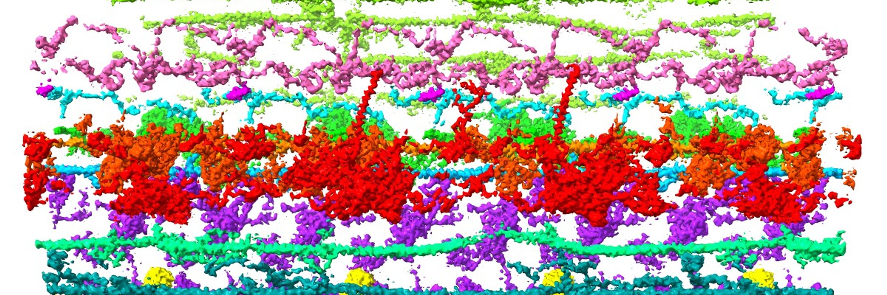
From reconstituted tomograms, dynein-2 molecules were found predominantly on the A-tubule side of the doublet microtubules, consistent with retrograde IFT trains in vivo.
5/8 🧵
5/8 🧵

January 28, 2025 at 7:05 PM
From reconstituted tomograms, dynein-2 molecules were found predominantly on the A-tubule side of the doublet microtubules, consistent with retrograde IFT trains in vivo.
5/8 🧵
5/8 🧵
To determine which side of the doublet microtubule dynein-2 binds to, we performed #cryoET aided by the Volta phase plate. Based on the morphology and distinctive microtubule inner proteins, the A- and B-tubules were identified.
4/8 🧵
4/8 🧵

January 28, 2025 at 7:05 PM
To determine which side of the doublet microtubule dynein-2 binds to, we performed #cryoET aided by the Volta phase plate. Based on the morphology and distinctive microtubule inner proteins, the A- and B-tubules were identified.
4/8 🧵
4/8 🧵
We performed in vitro reconstitution and #cryoEM observation, and found that dynein-2, the motor of the retrograde IFT, accumulated on one side of the doublet microtubule.
3/8 🧵
3/8 🧵

January 28, 2025 at 7:05 PM
We performed in vitro reconstitution and #cryoEM observation, and found that dynein-2, the motor of the retrograde IFT, accumulated on one side of the doublet microtubule.
3/8 🧵
3/8 🧵
Proud to be part of this paper published in @naturecomms.bsky.social. Using high-speed AFM, we have visualized the real-time translocation of substrate polypeptide by the single molecule of SecYEG-SecA!!!
Please check this amazing video taken by high-speed AFM!!!
www.nature.com/articles/s41...
Please check this amazing video taken by high-speed AFM!!!
www.nature.com/articles/s41...
January 8, 2025 at 12:43 PM
Proud to be part of this paper published in @naturecomms.bsky.social. Using high-speed AFM, we have visualized the real-time translocation of substrate polypeptide by the single molecule of SecYEG-SecA!!!
Please check this amazing video taken by high-speed AFM!!!
www.nature.com/articles/s41...
Please check this amazing video taken by high-speed AFM!!!
www.nature.com/articles/s41...
"The Cilia-Savvy Professor You Didn't Know You Needed" 😂
blueskyroast.com
blueskyroast.com

December 7, 2024 at 4:37 AM
"The Cilia-Savvy Professor You Didn't Know You Needed" 😂
blueskyroast.com
blueskyroast.com
I joined "The 47th Annual Meeting of the Molecular Biology Society of Japan" last week. Many interesting presentations, and I made new connections!

December 3, 2024 at 3:09 PM
I joined "The 47th Annual Meeting of the Molecular Biology Society of Japan" last week. Many interesting presentations, and I made new connections!
I already talked to some people, but as a member of Associate Editorial Board of Cytoskeleton Journal, I am putting together a special issue "Structures of microtubules and microtubule-related proteins". #microtubule #structure #cytoskeleton

November 26, 2024 at 12:51 PM
I already talked to some people, but as a member of Associate Editorial Board of Cytoskeleton Journal, I am putting together a special issue "Structures of microtubules and microtubule-related proteins". #microtubule #structure #cytoskeleton
Also, as a co-author, I helped analyze cryo-EM structures of P2X7 receptors with two antagonists. Panda🐼 P2X7 receptor was used. 😂
Reported as a reviewed preprint in eLife this year.
elifesciences.org/articles/92829
Reported as a reviewed preprint in eLife this year.
elifesciences.org/articles/92829

November 24, 2024 at 6:57 PM
Also, as a co-author, I helped analyze cryo-EM structures of P2X7 receptors with two antagonists. Panda🐼 P2X7 receptor was used. 😂
Reported as a reviewed preprint in eLife this year.
elifesciences.org/articles/92829
Reported as a reviewed preprint in eLife this year.
elifesciences.org/articles/92829
For #membraneprotein, a co-authored paper about cryo-EM structures of P2X4 receptors with two antagonists was published in Nature Communications in 2023. Insights into subtype-specific allosteric inhibition were obtained.
www.nature.com/articles/s41...
www.nature.com/articles/s41...

November 24, 2024 at 6:38 PM
For #membraneprotein, a co-authored paper about cryo-EM structures of P2X4 receptors with two antagonists was published in Nature Communications in 2023. Insights into subtype-specific allosteric inhibition were obtained.
www.nature.com/articles/s41...
www.nature.com/articles/s41...
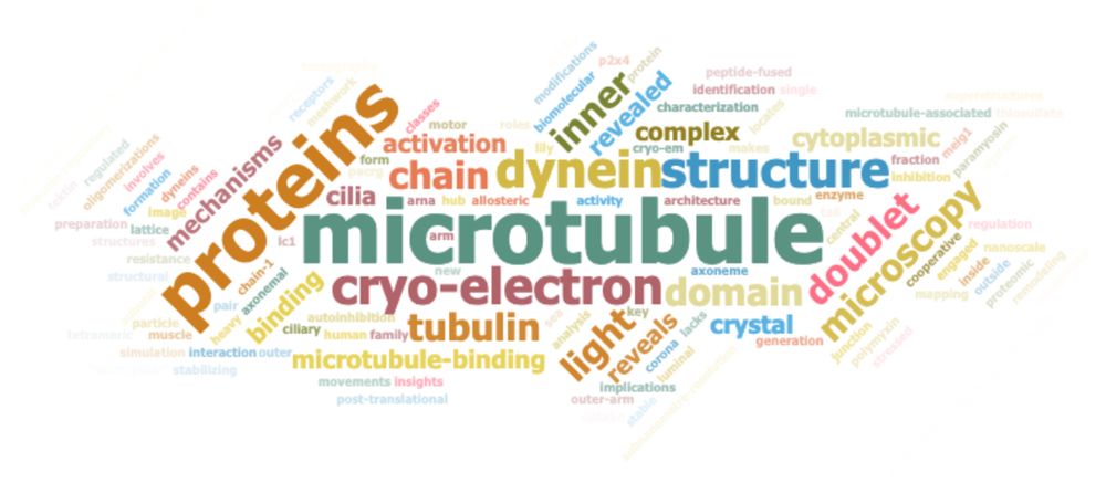
Made this with Scholar Goggler scholargoggler.com
@scholargoggler.bsky.social. I thought there were more cilia in the title, but not really. 😂
@scholargoggler.bsky.social. I thought there were more cilia in the title, but not really. 😂
November 24, 2024 at 12:42 PM
Made this with Scholar Goggler scholargoggler.com
@scholargoggler.bsky.social. I thought there were more cilia in the title, but not really. 😂
@scholargoggler.bsky.social. I thought there were more cilia in the title, but not really. 😂
Also, here's a different topic paper. In this paper, we characterized the properties of the biomolecular corona on nanoparticles by cryo-EM, cryo-ET, and simulation. I helped with the cryo-ET part. (Nature Communications, 2021)
www.nature.com/articles/s41...
www.nature.com/articles/s41...
November 24, 2024 at 8:47 AM
Also, here's a different topic paper. In this paper, we characterized the properties of the biomolecular corona on nanoparticles by cryo-EM, cryo-ET, and simulation. I helped with the cryo-ET part. (Nature Communications, 2021)
www.nature.com/articles/s41...
www.nature.com/articles/s41...
We also generated an exogenous protein with microtubule inside and outside binding sequences. Using this, we were able to reconstitute doublet microtubules and branching doublets in vitro, as confirmed by fluorescent and electron microscopies (Science Advances, 2022).
www.science.org/doi/full/10....
www.science.org/doi/full/10....

November 23, 2024 at 8:39 PM
We also generated an exogenous protein with microtubule inside and outside binding sequences. Using this, we were able to reconstitute doublet microtubules and branching doublets in vitro, as confirmed by fluorescent and electron microscopies (Science Advances, 2022).
www.science.org/doi/full/10....
www.science.org/doi/full/10....
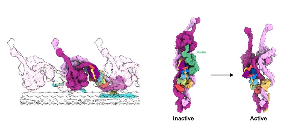
We also obtained the #cryo-EM structure of ODA bound to the doublet microtubule. By comparing active ODA structure with inactive ODA structure by Dr. Mali @dyneinassembly.bsky.social, we showed its conformational change upon activation (EMBO reports, 2021).
www.embopress.org/doi/full/10....
www.embopress.org/doi/full/10....
November 23, 2024 at 7:43 PM
We also obtained the #cryo-EM structure of ODA bound to the doublet microtubule. By comparing active ODA structure with inactive ODA structure by Dr. Mali @dyneinassembly.bsky.social, we showed its conformational change upon activation (EMBO reports, 2021).
www.embopress.org/doi/full/10....
www.embopress.org/doi/full/10....
For the outer dynein arm (ODA) of axonemal dynein, we showed that the light chain-1 (LC1) is located at the microtubule binding domain (MTBD) of the γ heavy chain. There are no other subunits binding MTBD, and it was an unexpected strategy of dynein regulation. www.molbiolcell.org/doi/full/10....

November 23, 2024 at 7:12 PM
For the outer dynein arm (ODA) of axonemal dynein, we showed that the light chain-1 (LC1) is located at the microtubule binding domain (MTBD) of the γ heavy chain. There are no other subunits binding MTBD, and it was an unexpected strategy of dynein regulation. www.molbiolcell.org/doi/full/10....
Ok, for the #dynein part!
This paper was published in @naturecellbiology.bsky.social in 2014, so definitely not recent, but we proposed that dynein-1's stacked conformation is autoinhibitory state for the first time. This model is widely accepted in the field now. www.nature.com/articles/ncb...
This paper was published in @naturecellbiology.bsky.social in 2014, so definitely not recent, but we proposed that dynein-1's stacked conformation is autoinhibitory state for the first time. This model is widely accepted in the field now. www.nature.com/articles/ncb...

November 23, 2024 at 6:58 PM
Ok, for the #dynein part!
This paper was published in @naturecellbiology.bsky.social in 2014, so definitely not recent, but we proposed that dynein-1's stacked conformation is autoinhibitory state for the first time. This model is widely accepted in the field now. www.nature.com/articles/ncb...
This paper was published in @naturecellbiology.bsky.social in 2014, so definitely not recent, but we proposed that dynein-1's stacked conformation is autoinhibitory state for the first time. This model is widely accepted in the field now. www.nature.com/articles/ncb...
Of course, there are other labs working on doublet microtubule structure by cryo-EM, and I had the pleasure of writing a Preview in Cell 'Tektin makes a microtubule a “micropillar”' about two papers showing mammalian doublet microtubules filled with tektins.
www.sciencedirect.com/science/arti...
www.sciencedirect.com/science/arti...

November 23, 2024 at 2:17 PM
Of course, there are other labs working on doublet microtubule structure by cryo-EM, and I had the pleasure of writing a Preview in Cell 'Tektin makes a microtubule a “micropillar”' about two papers showing mammalian doublet microtubules filled with tektins.
www.sciencedirect.com/science/arti...
www.sciencedirect.com/science/arti...
By combining #cryo-EM structure with mass spectrometry result, we identified proteins forming inner junction of doublet microtubule. From the model obtained, we presented a model of how B-tubule is tethered to the A-tubule. Published in @elife.bsky.social in 2020.
elifesciences.org/articles/52760
elifesciences.org/articles/52760
November 23, 2024 at 1:46 PM
By combining #cryo-EM structure with mass spectrometry result, we identified proteins forming inner junction of doublet microtubule. From the model obtained, we presented a model of how B-tubule is tethered to the A-tubule. Published in @elife.bsky.social in 2020.
elifesciences.org/articles/52760
elifesciences.org/articles/52760
In 2019, by improving the resolution of #cryo-EM structure of doublet microtubule from #cilia, we revealed the complex network of microtubule inner proteins (MIPs). Based on the structure, we presented a model of stabilization mechanism of doublet microtubules.
www.pnas.org/doi/abs/10.1...
www.pnas.org/doi/abs/10.1...

November 23, 2024 at 11:41 AM
In 2019, by improving the resolution of #cryo-EM structure of doublet microtubule from #cilia, we revealed the complex network of microtubule inner proteins (MIPs). Based on the structure, we presented a model of stabilization mechanism of doublet microtubules.
www.pnas.org/doi/abs/10.1...
www.pnas.org/doi/abs/10.1...
Following this, YeeD protein was identified as an essential component of YeeE in bacterial cells. YeeE-YeeD complex structure was obtained by X-ray crystallography. Based on other analysis results, YeeD receives and degrades the thiosulfate ions taken up by YeeE.
journals.plos.org/plosbiology/...
journals.plos.org/plosbiology/...

November 22, 2024 at 5:48 PM
Following this, YeeD protein was identified as an essential component of YeeE in bacterial cells. YeeE-YeeD complex structure was obtained by X-ray crystallography. Based on other analysis results, YeeD receives and degrades the thiosulfate ions taken up by YeeE.
journals.plos.org/plosbiology/...
journals.plos.org/plosbiology/...
In this paper published in Science Advances in 2020, we identified a membrane protein YeeE as a component of the novel #thiosulfate uptake pathway in bacteria. The structure of YeeE was obtained by X-ray crystallography.
www.science.org/doi/full/10....
www.science.org/doi/full/10....

November 22, 2024 at 5:29 PM
In this paper published in Science Advances in 2020, we identified a membrane protein YeeE as a component of the novel #thiosulfate uptake pathway in bacteria. The structure of YeeE was obtained by X-ray crystallography.
www.science.org/doi/full/10....
www.science.org/doi/full/10....
Co-author paper has been published in PNAS. Glad to contribute to the paper!
Cryo-EM structure of the zinc-activated channel (ZAC) in the Cys-loop receptor superfamily | PNAS
www.pnas.org/doi/10.1073/...
Cryo-EM structure of the zinc-activated channel (ZAC) in the Cys-loop receptor superfamily | PNAS
www.pnas.org/doi/10.1073/...

November 19, 2024 at 5:01 PM
Co-author paper has been published in PNAS. Glad to contribute to the paper!
Cryo-EM structure of the zinc-activated channel (ZAC) in the Cys-loop receptor superfamily | PNAS
www.pnas.org/doi/10.1073/...
Cryo-EM structure of the zinc-activated channel (ZAC) in the Cys-loop receptor superfamily | PNAS
www.pnas.org/doi/10.1073/...