


Electron Microscopy Facility, University of Oslo
@emlaboslo.bsky.social
Established in 1966, today equipped with 4 electron microscopes (two transmission electron microscopes (TEM) / two scanning electron microscopes (SEM)) and a wide range of preparation equipment covering most of the current preparation methods.
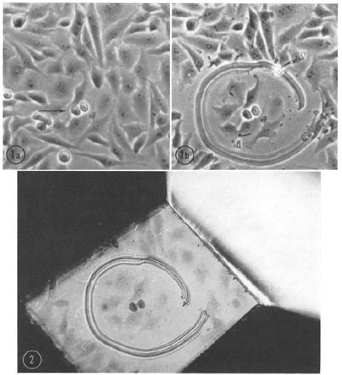
#Correlative Light and Electron Microscopy, #CLEM, is a modern #method combining light and #electronmicroscopy data. Although it is considered “new”, it was already used in the early 1960s, for instance to study the #ultrastructure of mitotic cells in this beautiful example: doi.org/10.1083/jcb....
January 13, 2025 at 7:47 AM
#Correlative Light and Electron Microscopy, #CLEM, is a modern #method combining light and #electronmicroscopy data. Although it is considered “new”, it was already used in the early 1960s, for instance to study the #ultrastructure of mitotic cells in this beautiful example: doi.org/10.1083/jcb....
This is probably what a #rotifer gets to see when it encounters a (seemingly bad-tempered) 6dpf #zebrafish larva. #Creepy! But maybe the fluffy #nostrils or the funny #neuromast beard make the situation a little better...
Scanning #electronmicroscopy / #SEM
Scanning #electronmicroscopy / #SEM

January 9, 2025 at 7:38 AM
This is probably what a #rotifer gets to see when it encounters a (seemingly bad-tempered) 6dpf #zebrafish larva. #Creepy! But maybe the fluffy #nostrils or the funny #neuromast beard make the situation a little better...
Scanning #electronmicroscopy / #SEM
Scanning #electronmicroscopy / #SEM
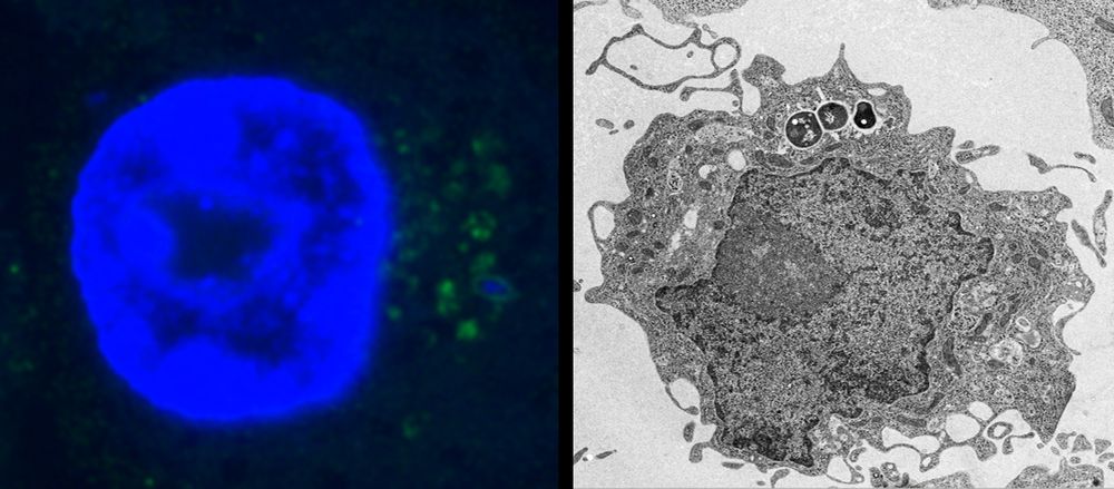
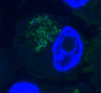
Still our favorite #review! Unfortunately, as relevant today as when it was published, it is an excellent illustration of the #problems arising from the neglect of #ultrastructure and #electronmicroscopy in #cell-biology and the over-reliance on #fluorescence #microscopy.
doi.org/10.1016/0962...
doi.org/10.1016/0962...
January 8, 2025 at 8:18 AM
Still our favorite #review! Unfortunately, as relevant today as when it was published, it is an excellent illustration of the #problems arising from the neglect of #ultrastructure and #electronmicroscopy in #cell-biology and the over-reliance on #fluorescence #microscopy.
doi.org/10.1016/0962...
doi.org/10.1016/0962...
Did you know that just 100-200 gold particles in your #immuno-EM data can give you an incredibly good idea of the #labelling distribution and reveal positive signals that you may not immediately recognise? It's amazing how reproducible this " #100-gold-method" is. #stereology
doi.org/10.1369/jhc....
doi.org/10.1369/jhc....

December 17, 2024 at 7:20 AM
Did you know that just 100-200 gold particles in your #immuno-EM data can give you an incredibly good idea of the #labelling distribution and reveal positive signals that you may not immediately recognise? It's amazing how reproducible this " #100-gold-method" is. #stereology
doi.org/10.1369/jhc....
doi.org/10.1369/jhc....
It's truly surprising that the #tokuyasu method is so underutilized in #fluorescence #microscopy! It eliminates the need for permeabilisation and increases Z resolution to 50nm on any microscope. It is also the method used in the Nobel Prize-winning work on #super-resolution.
doi.org/10.1177/0022...
doi.org/10.1177/0022...
December 13, 2024 at 7:30 AM
It's truly surprising that the #tokuyasu method is so underutilized in #fluorescence #microscopy! It eliminates the need for permeabilisation and increases Z resolution to 50nm on any microscope. It is also the method used in the Nobel Prize-winning work on #super-resolution.
doi.org/10.1177/0022...
doi.org/10.1177/0022...
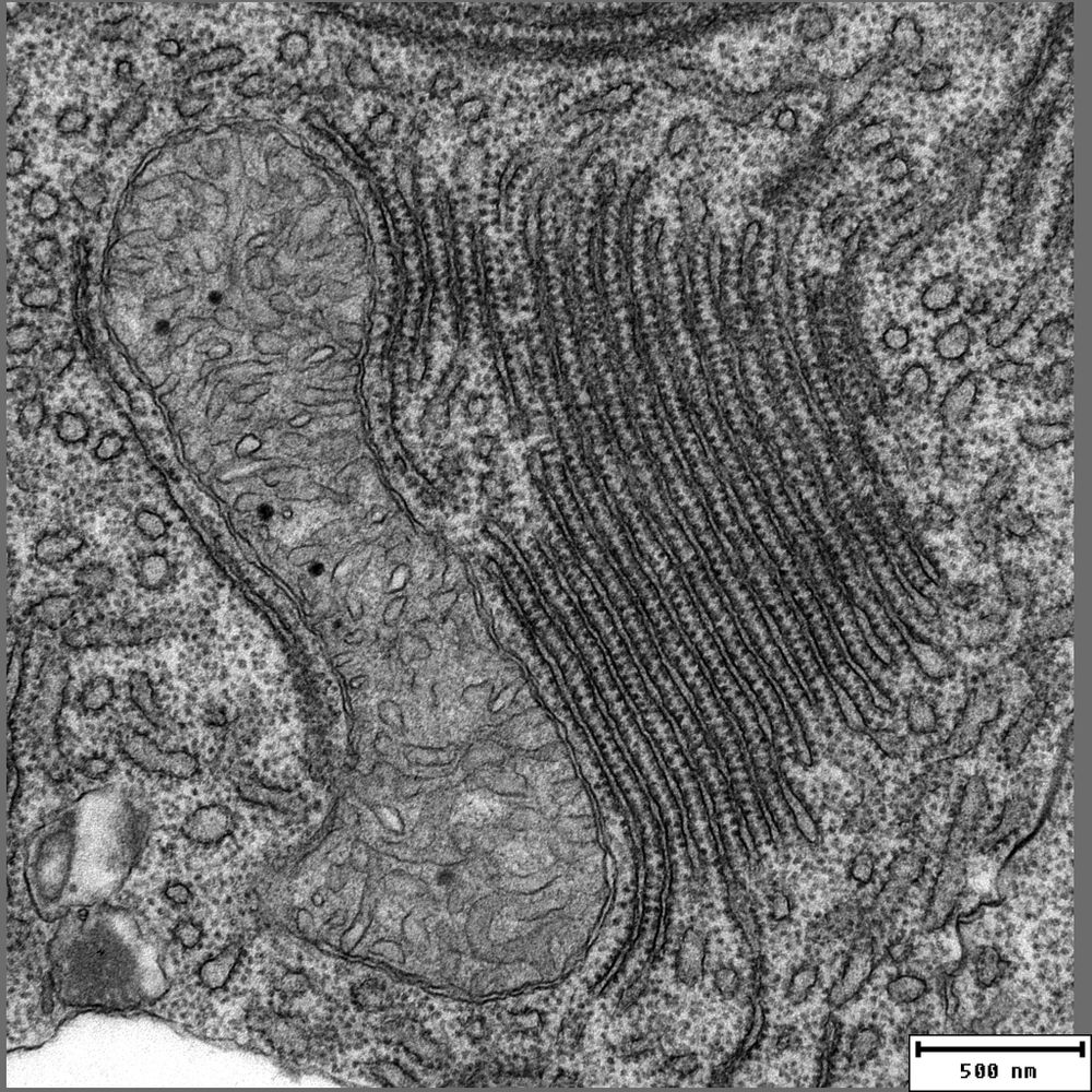
It is truly remarkable that Fritiof Sjöstrand was able to draw such precise conclusions about the cell in the era of primary osmium tetroxide #fixation, preceding the introduction of glutaraldehyde by Sabbatini in 1963. This 1956 review is a masterpiece of #ElectronMicroscopy
doi.org/10.1016/S007...
doi.org/10.1016/S007...

December 12, 2024 at 7:20 AM
It is truly remarkable that Fritiof Sjöstrand was able to draw such precise conclusions about the cell in the era of primary osmium tetroxide #fixation, preceding the introduction of glutaraldehyde by Sabbatini in 1963. This 1956 review is a masterpiece of #ElectronMicroscopy
doi.org/10.1016/S007...
doi.org/10.1016/S007...
A byproduct of a project on the #fish #immunesystem with Julien Resseguier from IBV @biovitenskap.bsky.social. The colored #SEM image shows a cross-section of the #gill #vasculature and the position of a #rodlet-cell (green) in the epithelium including its surface protrusion.
doi.org/10.1126/scia...
doi.org/10.1126/scia...

December 6, 2024 at 7:11 AM
A byproduct of a project on the #fish #immunesystem with Julien Resseguier from IBV @biovitenskap.bsky.social. The colored #SEM image shows a cross-section of the #gill #vasculature and the position of a #rodlet-cell (green) in the epithelium including its surface protrusion.
doi.org/10.1126/scia...
doi.org/10.1126/scia...
These hungry J774E #macrophages have probably overreached themselves a little with this aggregate of polystyrene #beads. We're not sure if teamwork- #phagocytosis is a promising approach for them. (false colored scanning electron #microscopy image)

November 28, 2024 at 8:31 PM
These hungry J774E #macrophages have probably overreached themselves a little with this aggregate of polystyrene #beads. We're not sure if teamwork- #phagocytosis is a promising approach for them. (false colored scanning electron #microscopy image)
#caveolae of the #zebrafish notochord. Whenever you come across them, you simply have to take an exposure. We haven't read up on them for a while, but we wonder whether their role here (apart from the mechanical) has been further worked out in the meantime -if there is one. doi.org/10.1016/j.cu...

November 28, 2024 at 9:44 AM
#caveolae of the #zebrafish notochord. Whenever you come across them, you simply have to take an exposure. We haven't read up on them for a while, but we wonder whether their role here (apart from the mechanical) has been further worked out in the meantime -if there is one. doi.org/10.1016/j.cu...
In case you feel like exploring #zebrafish #ultrastructure in the TEM but do not have an #electronmicroscope at hand, try one of our digital datasets. This is the pectoral fin of a 6dpf larva showing the mucosa through alcian blue. wohlmann.github.io/2024_EMMA_F0...

November 27, 2024 at 2:27 PM
In case you feel like exploring #zebrafish #ultrastructure in the TEM but do not have an #electronmicroscope at hand, try one of our digital datasets. This is the pectoral fin of a 6dpf larva showing the mucosa through alcian blue. wohlmann.github.io/2024_EMMA_F0...
Imagine a method capable of obtaining precise quantitative 3D information about a structure or molecular distribution from 2D sections in just a few minutes with only 200 simple counts. This almost magical method - ‘stereology’ - exists and is surprisingly underused. doi.org/10.1002/aja....

November 25, 2024 at 9:49 PM
Imagine a method capable of obtaining precise quantitative 3D information about a structure or molecular distribution from 2D sections in just a few minutes with only 200 simple counts. This almost magical method - ‘stereology’ - exists and is surprisingly underused. doi.org/10.1002/aja....