


Alexander Shakeel Bates
@asbates.bsky.social
Fly Neuroscientist and other good things.
The #BANC connectome sample was the best dissection of ~150 by @mindyisminsu.bsky.social! Behaviourally characterised by @debivort.bsky.social lab, microCT used to assess integrity before continuing. Then Minsu and @jasper-tms.bsky.social imaged over ~7 months
Details: github.com/jasper-tms/t...
Details: github.com/jasper-tms/t...

August 17, 2025 at 7:52 PM
The #BANC connectome sample was the best dissection of ~150 by @mindyisminsu.bsky.social! Behaviourally characterised by @debivort.bsky.social lab, microCT used to assess integrity before continuing. Then Minsu and @jasper-tms.bsky.social imaged over ~7 months
Details: github.com/jasper-tms/t...
Details: github.com/jasper-tms/t...
Explore neurons that enter/exit the fly central nervous system by body part with this clickable fly!: codex.flywire.ai/app/body_par...
(graphics: @mottcallie.bsky.social, tool: Arie Matisliah)
Vector graphics! github.com/wilson-lab/s...
Effector cells in 3D! ng.banc.community/2025a/effere...
(graphics: @mottcallie.bsky.social, tool: Arie Matisliah)
Vector graphics! github.com/wilson-lab/s...
Effector cells in 3D! ng.banc.community/2025a/effere...

August 7, 2025 at 10:33 PM
Explore neurons that enter/exit the fly central nervous system by body part with this clickable fly!: codex.flywire.ai/app/body_par...
(graphics: @mottcallie.bsky.social, tool: Arie Matisliah)
Vector graphics! github.com/wilson-lab/s...
Effector cells in 3D! ng.banc.community/2025a/effere...
(graphics: @mottcallie.bsky.social, tool: Arie Matisliah)
Vector graphics! github.com/wilson-lab/s...
Effector cells in 3D! ng.banc.community/2025a/effere...
For the BANC, @jasper-tms.bsky.social and I have added layers for neurons in other datasets to our neuroglancer environment. Search by name and see your favourite neurons in the same space! See an example here: ng.banc.community/2025a/DNa01-...

August 6, 2025 at 4:49 PM
For the BANC, @jasper-tms.bsky.social and I have added layers for neurons in other datasets to our neuroglancer environment. Search by name and see your favourite neurons in the same space! See an example here: ng.banc.community/2025a/DNa01-...
Public access to the first fly connectome that spans the whole CNS - BANC!: codex.flywire.ai?dataset=banc
Different from prior connectomes - it is brain + cord (think spinal cord)
We use it to ‘embody’ the system and find it resembles ‘subsumption architecture’ doi.org/10.1101/2025...
Different from prior connectomes - it is brain + cord (think spinal cord)
We use it to ‘embody’ the system and find it resembles ‘subsumption architecture’ doi.org/10.1101/2025...
August 2, 2025 at 2:30 PM
Public access to the first fly connectome that spans the whole CNS - BANC!: codex.flywire.ai?dataset=banc
Different from prior connectomes - it is brain + cord (think spinal cord)
We use it to ‘embody’ the system and find it resembles ‘subsumption architecture’ doi.org/10.1101/2025...
Different from prior connectomes - it is brain + cord (think spinal cord)
We use it to ‘embody’ the system and find it resembles ‘subsumption architecture’ doi.org/10.1101/2025...
Our fly brain connectome papers are now live (www.nature.com/immersive/d4...), and is getting traction (www.bbc.co.uk/news/article...). With ~139k neurons, >8k cell types, synapses annotated, transmitters predicted, ~134 evo-devo units denoted, so much is now possible in the fly.
October 3, 2024 at 12:26 AM
Our fly brain connectome papers are now live (www.nature.com/immersive/d4...), and is getting traction (www.bbc.co.uk/news/article...). With ~139k neurons, >8k cell types, synapses annotated, transmitters predicted, ~134 evo-devo units denoted, so much is now possible in the fly.
We hope these results will act as an accelerant for connectome-driven/informed hypotheses and neuroscientific investigation. We made these results available to the community as early as we could (#biorxiv @biorxiv-neursci.bsky.social in 2020). Our predictions have been used in >12 studies since.
May 19, 2024 at 2:22 AM
We hope these results will act as an accelerant for connectome-driven/informed hypotheses and neuroscientific investigation. We made these results available to the community as early as we could (#biorxiv @biorxiv-neursci.bsky.social in 2020). Our predictions have been used in >12 studies since.
But we feel our work is a big step forward. Our data (zenodo.org/records/1059...) and code (github.com/funkelab/syn...) are available. Results also available through Codex (codex.flywire.ai, FlyWire team) and we hope to see them in neuPrint (Janelia Fly EM) and on VirtualFlyBrain.org.
May 19, 2024 at 2:21 AM
But we feel our work is a big step forward. Our data (zenodo.org/records/1059...) and code (github.com/funkelab/syn...) are available. Results also available through Codex (codex.flywire.ai, FlyWire team) and we hope to see them in neuPrint (Janelia Fly EM) and on VirtualFlyBrain.org.
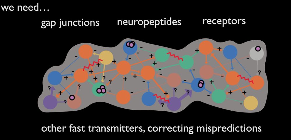
Not quite mission complete. We want more annotations. E.g. receptor localisation delineates excitatory from inhibitory glut connections, moving our +/ - labels to individual connections. Their annotation could enable ML to extract this molecular information from EM at scale.
May 19, 2024 at 2:21 AM
Not quite mission complete. We want more annotations. E.g. receptor localisation delineates excitatory from inhibitory glut connections, moving our +/ - labels to individual connections. Their annotation could enable ML to extract this molecular information from EM at scale.
Which proxy for synaptic signs!... With major assumptions, e.g. no co-transmission, and that glutamate is inhibitory - but, it seems ~80% of brain neurons may express both excitatory and inhibitory GluRs, and co-transmission esp. in monoaminergic neurons is reasonably common.
May 19, 2024 at 2:20 AM
Which proxy for synaptic signs!... With major assumptions, e.g. no co-transmission, and that glutamate is inhibitory - but, it seems ~80% of brain neurons may express both excitatory and inhibitory GluRs, and co-transmission esp. in monoaminergic neurons is reasonably common.
Which means, we had now ‘coloured in’ the connectome with transmitter labels…
May 19, 2024 at 2:20 AM
Which means, we had now ‘coloured in’ the connectome with transmitter labels…
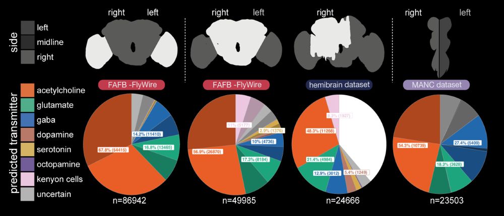
Yet overall, we had predictions which we felt were pretty good, across several data sets that together make up a whole (amalgam) Drosophila melanogaster nervous system! The neurotransmitter proportions turned out similar to those suggested by single-cell RNA-seq studies.
May 19, 2024 at 2:19 AM
Yet overall, we had predictions which we felt were pretty good, across several data sets that together make up a whole (amalgam) Drosophila melanogaster nervous system! The neurotransmitter proportions turned out similar to those suggested by single-cell RNA-seq studies.
In other cases, we see inconsistent results that look like genuine mispredictions, maybe <10% of our total neuronal predictions. The most obvious Kenyon cells, incorrectly predicted dopaminergic, rather than cholinergic. Our serotonin predictions are the least reliable.
May 19, 2024 at 2:19 AM
In other cases, we see inconsistent results that look like genuine mispredictions, maybe <10% of our total neuronal predictions. The most obvious Kenyon cells, incorrectly predicted dopaminergic, rather than cholinergic. Our serotonin predictions are the least reliable.
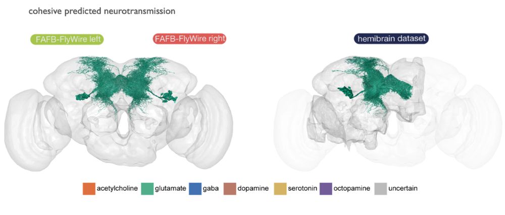
Here, for example, all members of a hemilineages are predicted glutamatergic (these are all tangential cells of the fan-shaped body).
May 19, 2024 at 2:18 AM
Here, for example, all members of a hemilineages are predicted glutamatergic (these are all tangential cells of the fan-shaped body).
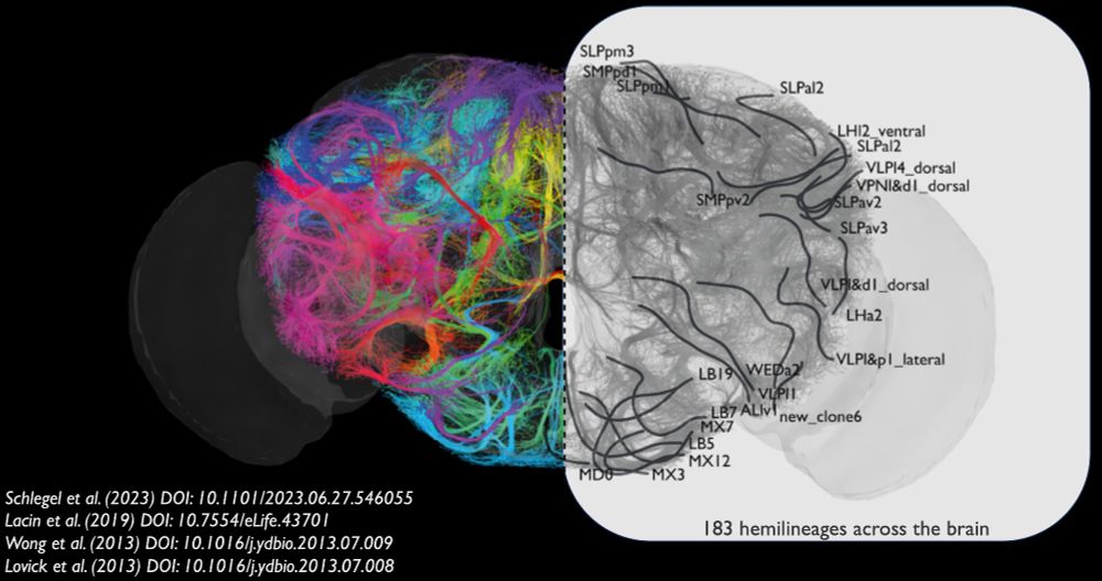
We also noticed higher-level principles. Lacin et al. (2019) had previously shown that neurons that are born together (in a ‘hemilineage’, ~100-200 neurons) express the same fast-acting transmitter in the ventral nerve cord. We found this held true in the ~183 brain hemilineages as well!
May 19, 2024 at 2:18 AM
We also noticed higher-level principles. Lacin et al. (2019) had previously shown that neurons that are born together (in a ‘hemilineage’, ~100-200 neurons) express the same fast-acting transmitter in the ventral nerve cord. We found this held true in the ~183 brain hemilineages as well!
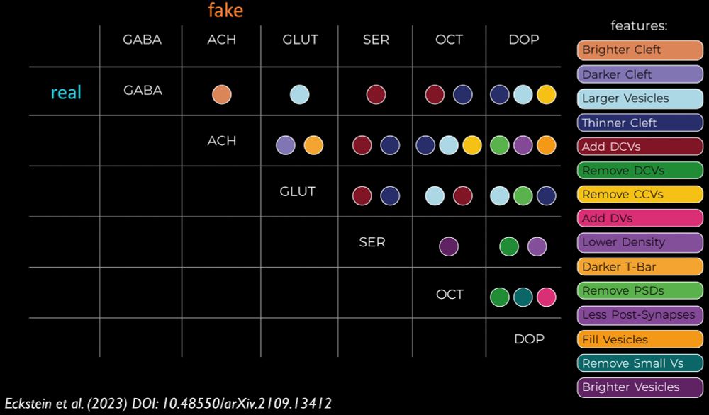
Now humans (i.e. Alicia Kun-Yang Lu, Thomson Rhymer, Samantha Finley-May, Tyler Paterson) could look at the images and work out what had been added to cause a ‘misprediction’. This matrix shows the features that will convert a real synapse (rows) into a different prediction by Synister (columns).
May 19, 2024 at 2:17 AM
Now humans (i.e. Alicia Kun-Yang Lu, Thomson Rhymer, Samantha Finley-May, Tyler Paterson) could look at the images and work out what had been added to cause a ‘misprediction’. This matrix shows the features that will convert a real synapse (rows) into a different prediction by Synister (columns).
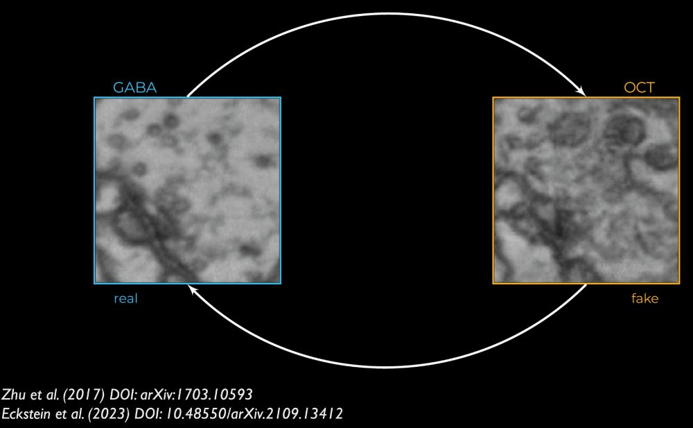
But how could it tell from a single synapse, what transmitter was present? Nils and Jan developed an explainable AI method (openreview.net/forum?id=Cn-...) to permute images of real synapses into counterfactuals that fool Synister, e.g. below, adding dark vesicle-like things throws prediction.
May 19, 2024 at 2:17 AM
But how could it tell from a single synapse, what transmitter was present? Nils and Jan developed an explainable AI method (openreview.net/forum?id=Cn-...) to permute images of real synapses into counterfactuals that fool Synister, e.g. below, adding dark vesicle-like things throws prediction.
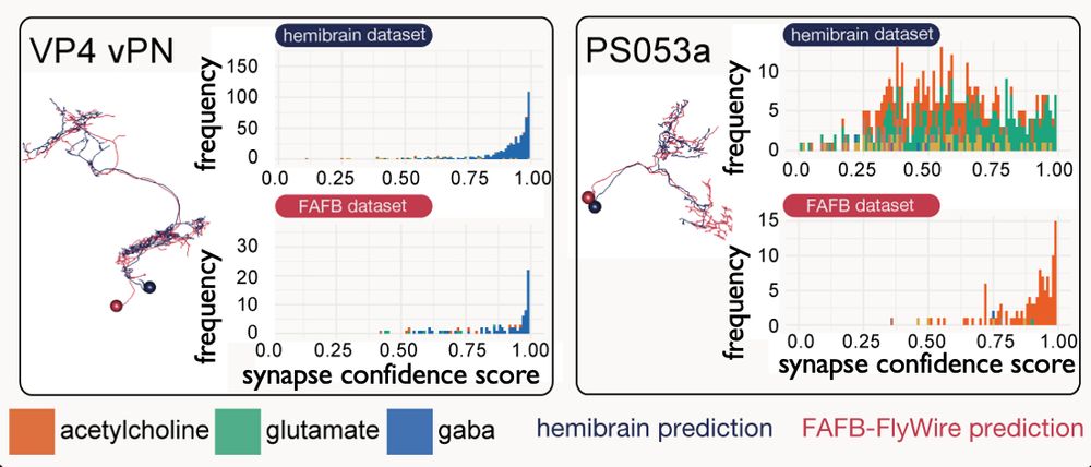
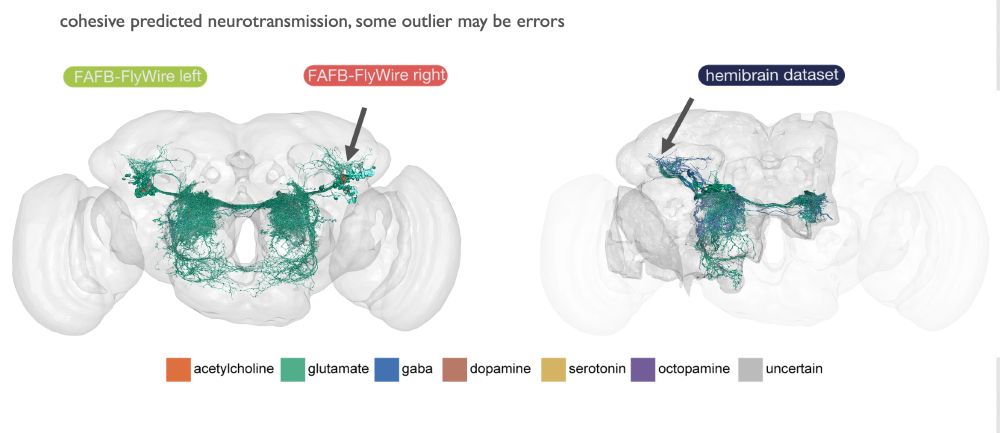
We also found that matched across datasets neurons get the same predictions, suggesting our predictions are varying with real biology, and are robust to inter-dataset or biological neuron origin differences.
May 19, 2024 at 2:16 AM
We also found that matched across datasets neurons get the same predictions, suggesting our predictions are varying with real biology, and are robust to inter-dataset or biological neuron origin differences.
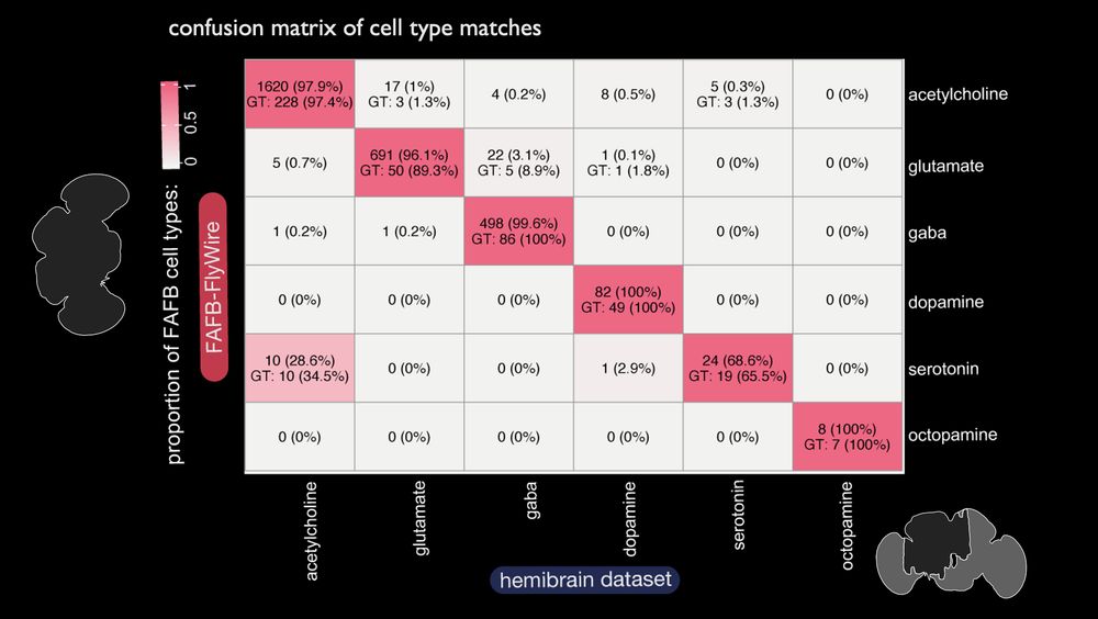
It worked remarkably well. Here, this confusion matrix shows that our prediction (columns) is correct (rows) at >80% rates for each transmitter class once we aggregate our per-synapse results by neuron. Overall, our accuracy was >90%.

May 19, 2024 at 2:15 AM
It worked remarkably well. Here, this confusion matrix shows that our prediction (columns) is correct (rows) at >80% rates for each transmitter class once we aggregate our per-synapse results by neuron. Overall, our accuracy was >90%.
But Nils Eckstein and Jan Funke were game. They developed ‘Synister’ ( github.com/funkelab/syn...) a deep convolutional neural network, which makes predictions from 640nm cubes, centred at presynaptic T-bars. Synapses had been manually annotated or automatically detected, e.g. Julia Buhmann et al.

May 19, 2024 at 2:14 AM
But Nils Eckstein and Jan Funke were game. They developed ‘Synister’ ( github.com/funkelab/syn...) a deep convolutional neural network, which makes predictions from 640nm cubes, centred at presynaptic T-bars. Synapses had been manually annotated or automatically detected, e.g. Julia Buhmann et al.
However, a priori we did not know if supervised learning for transmitter identity could work, because for electron micrographs of small invertebrates humans cannot see the difference between, say, the main excitatory transmitter (acetylcholine) and the main inhibitory one (GABA).

May 19, 2024 at 2:14 AM
However, a priori we did not know if supervised learning for transmitter identity could work, because for electron micrographs of small invertebrates humans cannot see the difference between, say, the main excitatory transmitter (acetylcholine) and the main inhibitory one (GABA).
In 2019, we only had ~300 so-identified cell types, ~3k neurons to learn with, from the Full Adult Fly Brain (FAFB) dataset manually made using CATMAID. From these, we got ~300k synapses for test/train. We assumed Dale’s law, that all synapses from the same neuron use the same transmitter.

May 19, 2024 at 2:14 AM
In 2019, we only had ~300 so-identified cell types, ~3k neurons to learn with, from the Full Adult Fly Brain (FAFB) dataset manually made using CATMAID. From these, we got ~300k synapses for test/train. We assumed Dale’s law, that all synapses from the same neuron use the same transmitter.
Stereotyped and unique neuron morphologies are the bridge that can link the EM connectome (right) to light microscopy research that has identified some neurotransmitter use using molecular biology (left), in order to build this ground truth.

May 19, 2024 at 2:10 AM
Stereotyped and unique neuron morphologies are the bridge that can link the EM connectome (right) to light microscopy research that has identified some neurotransmitter use using molecular biology (left), in order to build this ground truth.
After the Connectomics Meeting 2019, Jan Funke, Philipp Schlegel and I wondered if we could get at this by predicting neurotransmitter use across the ~50M synapses and ~130k neurons in the brain, by assembling ground truth for <1% of it: www.janelia.org/news/high-sc...

May 19, 2024 at 2:10 AM
After the Connectomics Meeting 2019, Jan Funke, Philipp Schlegel and I wondered if we could get at this by predicting neurotransmitter use across the ~50M synapses and ~130k neurons in the brain, by assembling ground truth for <1% of it: www.janelia.org/news/high-sc...
But we wanted to know this:

May 19, 2024 at 2:09 AM
But we wanted to know this:
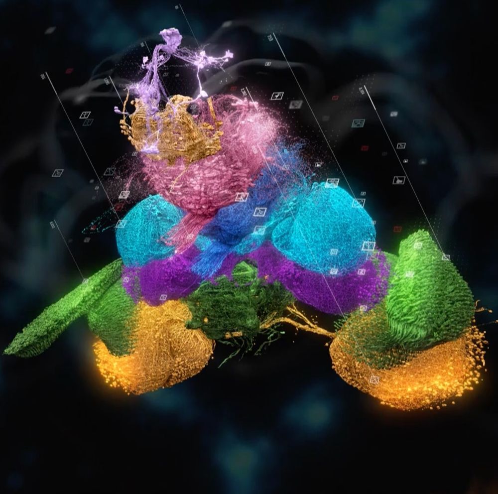
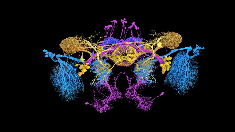
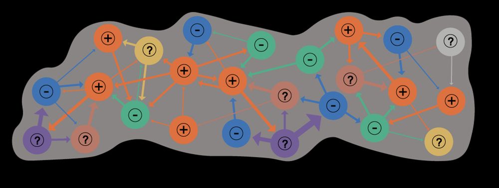
Put simply, recent work on the fly brain and ventral nerve cord (the invertebrate spinal cord analogue), has given us this for all bits of the CNS:

May 19, 2024 at 2:08 AM
Put simply, recent work on the fly brain and ventral nerve cord (the invertebrate spinal cord analogue), has given us this for all bits of the CNS: