


Developmental Biology
@devbiol.bsky.social
#DBfeature 🐠
A new powerful live imaging platform tracks Erk signalling dynamics in developing zebrafish hepatocytes at single-cell resolution.
By Faraz Ahmed Butt, Alessandro De Simone, Stefano Di Talia, Kenneth Poss
tinyurl.com/ve5vzp6c
A new powerful live imaging platform tracks Erk signalling dynamics in developing zebrafish hepatocytes at single-cell resolution.
By Faraz Ahmed Butt, Alessandro De Simone, Stefano Di Talia, Kenneth Poss
tinyurl.com/ve5vzp6c

November 25, 2025 at 12:35 PM
#DBfeature 🐠
A new powerful live imaging platform tracks Erk signalling dynamics in developing zebrafish hepatocytes at single-cell resolution.
By Faraz Ahmed Butt, Alessandro De Simone, Stefano Di Talia, Kenneth Poss
tinyurl.com/ve5vzp6c
A new powerful live imaging platform tracks Erk signalling dynamics in developing zebrafish hepatocytes at single-cell resolution.
By Faraz Ahmed Butt, Alessandro De Simone, Stefano Di Talia, Kenneth Poss
tinyurl.com/ve5vzp6c
#DBfeature 🧠🐣
Distinct functional domains of ASCL1 uniquely control neuronal differentiation and subtype identity, revealing the structural code behind neural fate decisions.
By Yuji Nakada, Madison Martinez, and Jane Johnson
tinyurl.com/3b6b325m
Distinct functional domains of ASCL1 uniquely control neuronal differentiation and subtype identity, revealing the structural code behind neural fate decisions.
By Yuji Nakada, Madison Martinez, and Jane Johnson
tinyurl.com/3b6b325m

November 21, 2025 at 2:20 PM
#DBfeature 🧠🐣
Distinct functional domains of ASCL1 uniquely control neuronal differentiation and subtype identity, revealing the structural code behind neural fate decisions.
By Yuji Nakada, Madison Martinez, and Jane Johnson
tinyurl.com/3b6b325m
Distinct functional domains of ASCL1 uniquely control neuronal differentiation and subtype identity, revealing the structural code behind neural fate decisions.
By Yuji Nakada, Madison Martinez, and Jane Johnson
tinyurl.com/3b6b325m
#DBfeature👁️
Shroom3 facilitates optic fissure closure via tissue alignment and reestablishment of apical-basal polarity during epithelial fusion
by Jessica Herstine, Jordyn Mensh, Timothy Plageman Jr. et al
www.sciencedirect.com/science/arti...
Shroom3 facilitates optic fissure closure via tissue alignment and reestablishment of apical-basal polarity during epithelial fusion
by Jessica Herstine, Jordyn Mensh, Timothy Plageman Jr. et al
www.sciencedirect.com/science/arti...

November 17, 2025 at 2:24 PM
#DBfeature👁️
Shroom3 facilitates optic fissure closure via tissue alignment and reestablishment of apical-basal polarity during epithelial fusion
by Jessica Herstine, Jordyn Mensh, Timothy Plageman Jr. et al
www.sciencedirect.com/science/arti...
Shroom3 facilitates optic fissure closure via tissue alignment and reestablishment of apical-basal polarity during epithelial fusion
by Jessica Herstine, Jordyn Mensh, Timothy Plageman Jr. et al
www.sciencedirect.com/science/arti...
#DBfeature
Teaching developmental biology through a biocultural lens can challenge biological essentialism and build a more equitable science
By Julia Paxson
tinyurl.com/43htbxfx
#SpecialIssue in Teaching #DevBio for Social Change
Teaching developmental biology through a biocultural lens can challenge biological essentialism and build a more equitable science
By Julia Paxson
tinyurl.com/43htbxfx
#SpecialIssue in Teaching #DevBio for Social Change
November 14, 2025 at 2:43 PM
#DBfeature
Teaching developmental biology through a biocultural lens can challenge biological essentialism and build a more equitable science
By Julia Paxson
tinyurl.com/43htbxfx
#SpecialIssue in Teaching #DevBio for Social Change
Teaching developmental biology through a biocultural lens can challenge biological essentialism and build a more equitable science
By Julia Paxson
tinyurl.com/43htbxfx
#SpecialIssue in Teaching #DevBio for Social Change
#DBfeature
Multidisciplinary educational approach to reproductive technology links developmental biology, ethics, and law to prepare biologists for real-world controversies
By J Azzi, Z Wehbi, P Hussein Kobeissy, R Kerek
tinyurl.com/2xvm64ue
#SpecialIssue in Teaching #DevBio for Social Change
Multidisciplinary educational approach to reproductive technology links developmental biology, ethics, and law to prepare biologists for real-world controversies
By J Azzi, Z Wehbi, P Hussein Kobeissy, R Kerek
tinyurl.com/2xvm64ue
#SpecialIssue in Teaching #DevBio for Social Change

November 12, 2025 at 1:51 PM
#DBfeature
Multidisciplinary educational approach to reproductive technology links developmental biology, ethics, and law to prepare biologists for real-world controversies
By J Azzi, Z Wehbi, P Hussein Kobeissy, R Kerek
tinyurl.com/2xvm64ue
#SpecialIssue in Teaching #DevBio for Social Change
Multidisciplinary educational approach to reproductive technology links developmental biology, ethics, and law to prepare biologists for real-world controversies
By J Azzi, Z Wehbi, P Hussein Kobeissy, R Kerek
tinyurl.com/2xvm64ue
#SpecialIssue in Teaching #DevBio for Social Change
#DBfeature
Fritz Müller’s "Für Darwin" (1864) bridged evolution and development, anticipating evo-devo and warning to the dangers of scientific dogma
By Scott Gilbert and Beatrice Steinert
tinyurl.com/3sccvcr5
#SpecialIssue on Research that transformed #DevBio
Fritz Müller’s "Für Darwin" (1864) bridged evolution and development, anticipating evo-devo and warning to the dangers of scientific dogma
By Scott Gilbert and Beatrice Steinert
tinyurl.com/3sccvcr5
#SpecialIssue on Research that transformed #DevBio

November 10, 2025 at 12:35 PM
#DBfeature
Fritz Müller’s "Für Darwin" (1864) bridged evolution and development, anticipating evo-devo and warning to the dangers of scientific dogma
By Scott Gilbert and Beatrice Steinert
tinyurl.com/3sccvcr5
#SpecialIssue on Research that transformed #DevBio
Fritz Müller’s "Für Darwin" (1864) bridged evolution and development, anticipating evo-devo and warning to the dangers of scientific dogma
By Scott Gilbert and Beatrice Steinert
tinyurl.com/3sccvcr5
#SpecialIssue on Research that transformed #DevBio
#DBfeature
Developmental life history transitions can be shaped by structural inequities: Insights from the sociology of race
by Sarah McMenamin, Latrica Best
sciencedirect.com/science/arti...
Developmental life history transitions can be shaped by structural inequities: Insights from the sociology of race
by Sarah McMenamin, Latrica Best
sciencedirect.com/science/arti...

November 7, 2025 at 10:25 PM
#DBfeature
Developmental life history transitions can be shaped by structural inequities: Insights from the sociology of race
by Sarah McMenamin, Latrica Best
sciencedirect.com/science/arti...
Developmental life history transitions can be shaped by structural inequities: Insights from the sociology of race
by Sarah McMenamin, Latrica Best
sciencedirect.com/science/arti...
#DBfeature 🐣🐠
Cereblon E3 ligase complex genes are expressed in tissues sensitive to thalidomide in chicken and zebrafish embryos but are unchanged following thalidomide exposure
By LR Fraga, J Reeves, C Mahony, L Erskine, N Vargesson
tinyurl.com/2fx6pbay
#SpecialIssue in Avian Model Systems
Cereblon E3 ligase complex genes are expressed in tissues sensitive to thalidomide in chicken and zebrafish embryos but are unchanged following thalidomide exposure
By LR Fraga, J Reeves, C Mahony, L Erskine, N Vargesson
tinyurl.com/2fx6pbay
#SpecialIssue in Avian Model Systems

November 6, 2025 at 2:56 PM
#DBfeature 🐣🐠
Cereblon E3 ligase complex genes are expressed in tissues sensitive to thalidomide in chicken and zebrafish embryos but are unchanged following thalidomide exposure
By LR Fraga, J Reeves, C Mahony, L Erskine, N Vargesson
tinyurl.com/2fx6pbay
#SpecialIssue in Avian Model Systems
Cereblon E3 ligase complex genes are expressed in tissues sensitive to thalidomide in chicken and zebrafish embryos but are unchanged following thalidomide exposure
By LR Fraga, J Reeves, C Mahony, L Erskine, N Vargesson
tinyurl.com/2fx6pbay
#SpecialIssue in Avian Model Systems
#DBfeature 🐦🦴
Micro-CT 3D structures provide new criteria which can be used to identify medullary bone and female individuals in the avian fossil record.
By Crane AH, Baldry CJ, Rankin KE, Clarkin CE, Williams KA, Gostling NJ.
tinyurl.com/pw5s23js
#SpecialIssue on Avian model systems
Micro-CT 3D structures provide new criteria which can be used to identify medullary bone and female individuals in the avian fossil record.
By Crane AH, Baldry CJ, Rankin KE, Clarkin CE, Williams KA, Gostling NJ.
tinyurl.com/pw5s23js
#SpecialIssue on Avian model systems

November 5, 2025 at 1:38 PM
#DBfeature 🐦🦴
Micro-CT 3D structures provide new criteria which can be used to identify medullary bone and female individuals in the avian fossil record.
By Crane AH, Baldry CJ, Rankin KE, Clarkin CE, Williams KA, Gostling NJ.
tinyurl.com/pw5s23js
#SpecialIssue on Avian model systems
Micro-CT 3D structures provide new criteria which can be used to identify medullary bone and female individuals in the avian fossil record.
By Crane AH, Baldry CJ, Rankin KE, Clarkin CE, Williams KA, Gostling NJ.
tinyurl.com/pw5s23js
#SpecialIssue on Avian model systems
#DBfeature #Review
This review paper elucidates the organizational principles underlying the integument.
By Chuong, C. M., Wu, P., Yu, Z., Liang, Y. C., & Widelitz, R. B.
doi.org/10.1016/j.yd...
This review paper elucidates the organizational principles underlying the integument.
By Chuong, C. M., Wu, P., Yu, Z., Liang, Y. C., & Widelitz, R. B.
doi.org/10.1016/j.yd...

November 4, 2025 at 10:11 AM
#DBfeature #Review
This review paper elucidates the organizational principles underlying the integument.
By Chuong, C. M., Wu, P., Yu, Z., Liang, Y. C., & Widelitz, R. B.
doi.org/10.1016/j.yd...
This review paper elucidates the organizational principles underlying the integument.
By Chuong, C. M., Wu, P., Yu, Z., Liang, Y. C., & Widelitz, R. B.
doi.org/10.1016/j.yd...
#DBfeature Review
Cyclic renewal in three ectodermal appendage follicles: Hairs, feathers and teeth
by Ping Wu et al
www.sciencedirect.com/science/arti...
Cyclic renewal in three ectodermal appendage follicles: Hairs, feathers and teeth
by Ping Wu et al
www.sciencedirect.com/science/arti...

November 3, 2025 at 2:22 PM
#DBfeature Review
Cyclic renewal in three ectodermal appendage follicles: Hairs, feathers and teeth
by Ping Wu et al
www.sciencedirect.com/science/arti...
Cyclic renewal in three ectodermal appendage follicles: Hairs, feathers and teeth
by Ping Wu et al
www.sciencedirect.com/science/arti...
#DBfeature 🪰
Drosophila grh relies on multiple enhancers for robust expression in neural stem cells, with different combinations playing a critical role in regulating its expression in subsets of these cells
By R Sipani, Y Rawal, J Barman, P Abbur, V Kurlawala, & R Joshi
tinyurl.com/2aufrc2w
Drosophila grh relies on multiple enhancers for robust expression in neural stem cells, with different combinations playing a critical role in regulating its expression in subsets of these cells
By R Sipani, Y Rawal, J Barman, P Abbur, V Kurlawala, & R Joshi
tinyurl.com/2aufrc2w

October 31, 2025 at 2:51 PM
#DBfeature 🪰
Drosophila grh relies on multiple enhancers for robust expression in neural stem cells, with different combinations playing a critical role in regulating its expression in subsets of these cells
By R Sipani, Y Rawal, J Barman, P Abbur, V Kurlawala, & R Joshi
tinyurl.com/2aufrc2w
Drosophila grh relies on multiple enhancers for robust expression in neural stem cells, with different combinations playing a critical role in regulating its expression in subsets of these cells
By R Sipani, Y Rawal, J Barman, P Abbur, V Kurlawala, & R Joshi
tinyurl.com/2aufrc2w
#DBfeature 🐟
Retinoic acid promotes second heart field addition & regulates ventral aorta patterning in zebrafish.
-by Austin Griffin, Allison Small, Jennifer Schumacher, et al
www.sciencedirect.com/science/arti...
Retinoic acid promotes second heart field addition & regulates ventral aorta patterning in zebrafish.
-by Austin Griffin, Allison Small, Jennifer Schumacher, et al
www.sciencedirect.com/science/arti...

October 30, 2025 at 3:02 PM
#DBfeature 🐟
Retinoic acid promotes second heart field addition & regulates ventral aorta patterning in zebrafish.
-by Austin Griffin, Allison Small, Jennifer Schumacher, et al
www.sciencedirect.com/science/arti...
Retinoic acid promotes second heart field addition & regulates ventral aorta patterning in zebrafish.
-by Austin Griffin, Allison Small, Jennifer Schumacher, et al
www.sciencedirect.com/science/arti...
#DBfeature 🐭
Wnt3a plays an important role in Wolffian duct development by regulating apicobasal polarity.
By Hayashi, S., Suzuki, H., Takada, S., & Takemoto, T.
doi.org/10.1016/j.yd...
Wnt3a plays an important role in Wolffian duct development by regulating apicobasal polarity.
By Hayashi, S., Suzuki, H., Takada, S., & Takemoto, T.
doi.org/10.1016/j.yd...

October 29, 2025 at 6:49 PM
#DBfeature 🐭
Wnt3a plays an important role in Wolffian duct development by regulating apicobasal polarity.
By Hayashi, S., Suzuki, H., Takada, S., & Takemoto, T.
doi.org/10.1016/j.yd...
Wnt3a plays an important role in Wolffian duct development by regulating apicobasal polarity.
By Hayashi, S., Suzuki, H., Takada, S., & Takemoto, T.
doi.org/10.1016/j.yd...
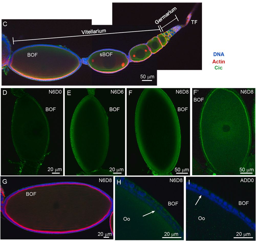
#DBfeature 🪳
The transcription factor Capicua maintains the oocyte polarity in the panoistic ovary of the German cockroach
"Cic interacts with Notch and EGFR pathways"
by Nashwa Elshaer, Jorge Escudero, Maria-Dolors Piulachs
www.sciencedirect.com/science/arti...
The transcription factor Capicua maintains the oocyte polarity in the panoistic ovary of the German cockroach
"Cic interacts with Notch and EGFR pathways"
by Nashwa Elshaer, Jorge Escudero, Maria-Dolors Piulachs
www.sciencedirect.com/science/arti...
October 28, 2025 at 1:47 PM
#DBfeature 🪳
The transcription factor Capicua maintains the oocyte polarity in the panoistic ovary of the German cockroach
"Cic interacts with Notch and EGFR pathways"
by Nashwa Elshaer, Jorge Escudero, Maria-Dolors Piulachs
www.sciencedirect.com/science/arti...
The transcription factor Capicua maintains the oocyte polarity in the panoistic ovary of the German cockroach
"Cic interacts with Notch and EGFR pathways"
by Nashwa Elshaer, Jorge Escudero, Maria-Dolors Piulachs
www.sciencedirect.com/science/arti...
#DBfeature
Scientists have identified a provisional regulatory circuit downstream of the transcription factor Pax3/7 operating in the descending decussating neurons (ddNs) of the tunicate Ciona robusta.
By Kim, K., Piekarz, K. M., & Stolfi, A.
doi.org/10.1016/j.yd...
Scientists have identified a provisional regulatory circuit downstream of the transcription factor Pax3/7 operating in the descending decussating neurons (ddNs) of the tunicate Ciona robusta.
By Kim, K., Piekarz, K. M., & Stolfi, A.
doi.org/10.1016/j.yd...

October 27, 2025 at 5:04 PM
#DBfeature
Scientists have identified a provisional regulatory circuit downstream of the transcription factor Pax3/7 operating in the descending decussating neurons (ddNs) of the tunicate Ciona robusta.
By Kim, K., Piekarz, K. M., & Stolfi, A.
doi.org/10.1016/j.yd...
Scientists have identified a provisional regulatory circuit downstream of the transcription factor Pax3/7 operating in the descending decussating neurons (ddNs) of the tunicate Ciona robusta.
By Kim, K., Piekarz, K. M., & Stolfi, A.
doi.org/10.1016/j.yd...
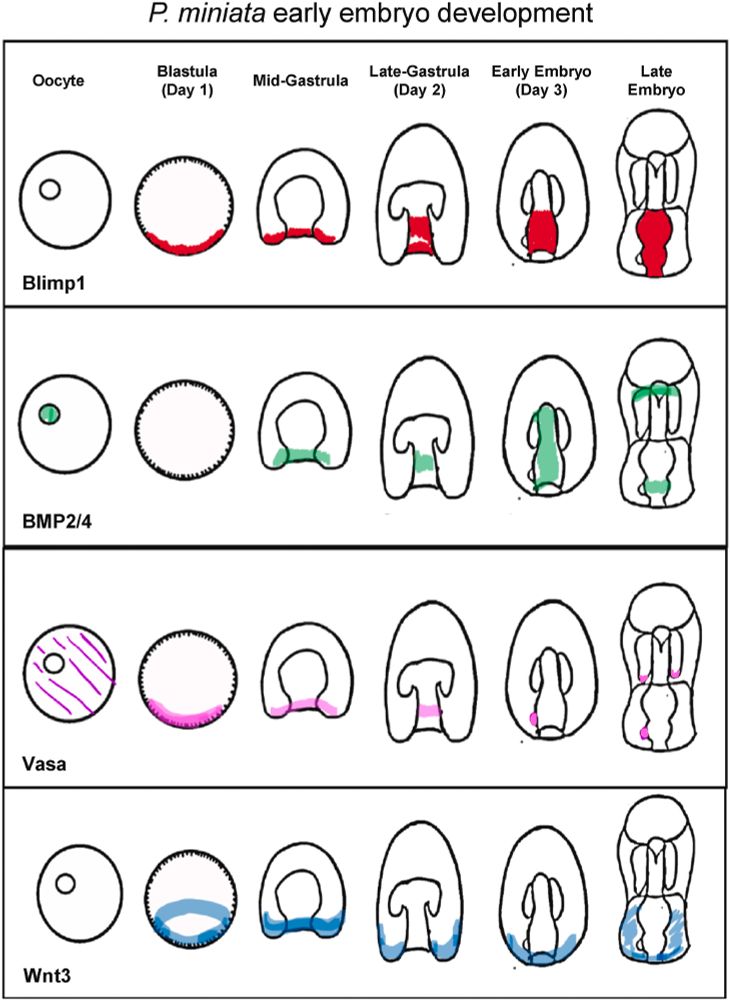
#DBfeature #EvoDevo
mRNA splicing variants of the transcription factor Blimp1 differentially regulate germline genes in echinoderms
"Each Blimp1 isoform has distinct functions within & between species"
by Gerardo Reyes, Nathalie Oulhen, Gary Wessel
www.sciencedirect.com/science/arti...
mRNA splicing variants of the transcription factor Blimp1 differentially regulate germline genes in echinoderms
"Each Blimp1 isoform has distinct functions within & between species"
by Gerardo Reyes, Nathalie Oulhen, Gary Wessel
www.sciencedirect.com/science/arti...
October 24, 2025 at 12:53 PM
#DBfeature #EvoDevo
mRNA splicing variants of the transcription factor Blimp1 differentially regulate germline genes in echinoderms
"Each Blimp1 isoform has distinct functions within & between species"
by Gerardo Reyes, Nathalie Oulhen, Gary Wessel
www.sciencedirect.com/science/arti...
mRNA splicing variants of the transcription factor Blimp1 differentially regulate germline genes in echinoderms
"Each Blimp1 isoform has distinct functions within & between species"
by Gerardo Reyes, Nathalie Oulhen, Gary Wessel
www.sciencedirect.com/science/arti...
#DBfeature 🧑🔬
David Sherwood draws a powerful parallel between morphogenesis and a scientific career, showing how becoming a biologist is a continual process of shaping, adaptation, and growth. Like development, a scientific life unfolds through adaptation and emergence.
tinyurl.com/6yd46wcx
David Sherwood draws a powerful parallel between morphogenesis and a scientific career, showing how becoming a biologist is a continual process of shaping, adaptation, and growth. Like development, a scientific life unfolds through adaptation and emergence.
tinyurl.com/6yd46wcx

October 23, 2025 at 12:42 PM
#DBfeature 🧑🔬
David Sherwood draws a powerful parallel between morphogenesis and a scientific career, showing how becoming a biologist is a continual process of shaping, adaptation, and growth. Like development, a scientific life unfolds through adaptation and emergence.
tinyurl.com/6yd46wcx
David Sherwood draws a powerful parallel between morphogenesis and a scientific career, showing how becoming a biologist is a continual process of shaping, adaptation, and growth. Like development, a scientific life unfolds through adaptation and emergence.
tinyurl.com/6yd46wcx
#DBfeature Review 🪽
Feather Morphogenesis
Keratinization and cornification of avian skin appendages during development. Insights from immunolabeling and electron microscopic studies
by Lorenzo Alibardi
www.sciencedirect.com/science/arti...
Feather Morphogenesis
Keratinization and cornification of avian skin appendages during development. Insights from immunolabeling and electron microscopic studies
by Lorenzo Alibardi
www.sciencedirect.com/science/arti...

October 22, 2025 at 2:21 PM
#DBfeature Review 🪽
Feather Morphogenesis
Keratinization and cornification of avian skin appendages during development. Insights from immunolabeling and electron microscopic studies
by Lorenzo Alibardi
www.sciencedirect.com/science/arti...
Feather Morphogenesis
Keratinization and cornification of avian skin appendages during development. Insights from immunolabeling and electron microscopic studies
by Lorenzo Alibardi
www.sciencedirect.com/science/arti...
#DBfeature
Building a genetic map of limb development driven by the apical ectodermal ridge
By Lee Niswander
tinyurl.com/bdzrjsyz
#SpecialIssue on Research that Transformed #DevelopmentalBiology
Building a genetic map of limb development driven by the apical ectodermal ridge
By Lee Niswander
tinyurl.com/bdzrjsyz
#SpecialIssue on Research that Transformed #DevelopmentalBiology

October 20, 2025 at 12:02 PM
#DBfeature
Building a genetic map of limb development driven by the apical ectodermal ridge
By Lee Niswander
tinyurl.com/bdzrjsyz
#SpecialIssue on Research that Transformed #DevelopmentalBiology
Building a genetic map of limb development driven by the apical ectodermal ridge
By Lee Niswander
tinyurl.com/bdzrjsyz
#SpecialIssue on Research that Transformed #DevelopmentalBiology
DBfeature 🌸
How homeotic mutants shaped the ABC model, transforming our understanding of floral patterning
By Zhongchi Liu
tinyurl.com/2tc58fbs
#SpecialIssue on Research that Transformed #DevelopmentalBiology
How homeotic mutants shaped the ABC model, transforming our understanding of floral patterning
By Zhongchi Liu
tinyurl.com/2tc58fbs
#SpecialIssue on Research that Transformed #DevelopmentalBiology

October 17, 2025 at 3:36 PM
DBfeature 🌸
How homeotic mutants shaped the ABC model, transforming our understanding of floral patterning
By Zhongchi Liu
tinyurl.com/2tc58fbs
#SpecialIssue on Research that Transformed #DevelopmentalBiology
How homeotic mutants shaped the ABC model, transforming our understanding of floral patterning
By Zhongchi Liu
tinyurl.com/2tc58fbs
#SpecialIssue on Research that Transformed #DevelopmentalBiology
#DBfeature
From peas to animal embryogenesis: how pioneering work in genetics shaped our understanding of development
By Abraham Fainsod and Martin Blum
tinyurl.com/5n8h423x
#SpecialIssue on Research that Transformed #DevelopmentalBiology
From peas to animal embryogenesis: how pioneering work in genetics shaped our understanding of development
By Abraham Fainsod and Martin Blum
tinyurl.com/5n8h423x
#SpecialIssue on Research that Transformed #DevelopmentalBiology

October 16, 2025 at 2:22 PM
#DBfeature
From peas to animal embryogenesis: how pioneering work in genetics shaped our understanding of development
By Abraham Fainsod and Martin Blum
tinyurl.com/5n8h423x
#SpecialIssue on Research that Transformed #DevelopmentalBiology
From peas to animal embryogenesis: how pioneering work in genetics shaped our understanding of development
By Abraham Fainsod and Martin Blum
tinyurl.com/5n8h423x
#SpecialIssue on Research that Transformed #DevelopmentalBiology
#DBfeature 🐣
PRDM14 is essential for vertebrate gastrulation and safeguards avian germ cell identity by antagonising FGF-induced differentiation
By D Doddamani, D Carlson, L McTeir, L Taylor, S Nandi, M Davey, M McGrew, J Glover
tinyurl.com/2wpakkyd
#SpecialIssue on Avian Model Systems
PRDM14 is essential for vertebrate gastrulation and safeguards avian germ cell identity by antagonising FGF-induced differentiation
By D Doddamani, D Carlson, L McTeir, L Taylor, S Nandi, M Davey, M McGrew, J Glover
tinyurl.com/2wpakkyd
#SpecialIssue on Avian Model Systems

October 15, 2025 at 1:08 PM
#DBfeature 🐣
PRDM14 is essential for vertebrate gastrulation and safeguards avian germ cell identity by antagonising FGF-induced differentiation
By D Doddamani, D Carlson, L McTeir, L Taylor, S Nandi, M Davey, M McGrew, J Glover
tinyurl.com/2wpakkyd
#SpecialIssue on Avian Model Systems
PRDM14 is essential for vertebrate gastrulation and safeguards avian germ cell identity by antagonising FGF-induced differentiation
By D Doddamani, D Carlson, L McTeir, L Taylor, S Nandi, M Davey, M McGrew, J Glover
tinyurl.com/2wpakkyd
#SpecialIssue on Avian Model Systems
#DBfeature
Micro-CT 3D structures provide new criteria can be used to identify medullary bone and female individuals in the avian fossil record.
Crane A H @gruiformabi.bsky.social, Baldry C J, Rankin K E, Clarkin C E @clarkinbonebiol.bsky.social, Williams K A, Gostling N J @neilgostling.bsky.social
Micro-CT 3D structures provide new criteria can be used to identify medullary bone and female individuals in the avian fossil record.
Crane A H @gruiformabi.bsky.social, Baldry C J, Rankin K E, Clarkin C E @clarkinbonebiol.bsky.social, Williams K A, Gostling N J @neilgostling.bsky.social
October 14, 2025 at 1:27 PM
#DBfeature
Micro-CT 3D structures provide new criteria can be used to identify medullary bone and female individuals in the avian fossil record.
Crane A H @gruiformabi.bsky.social, Baldry C J, Rankin K E, Clarkin C E @clarkinbonebiol.bsky.social, Williams K A, Gostling N J @neilgostling.bsky.social
Micro-CT 3D structures provide new criteria can be used to identify medullary bone and female individuals in the avian fossil record.
Crane A H @gruiformabi.bsky.social, Baldry C J, Rankin K E, Clarkin C E @clarkinbonebiol.bsky.social, Williams K A, Gostling N J @neilgostling.bsky.social
#DBfeature 🐣
The chicken embryo brings new insights into the tissue specific evolutionary role of WFDC1 during amniote development
-T Metzker-Pinto, YTH Tran, I Buzzatto-Leite, L Lok, JF Sampar, HF Carvalho, G del Monte-Nieto, LE Alvares
tinyurl.com/3abhm3d5
#SpecialIssue on Avian model systems
The chicken embryo brings new insights into the tissue specific evolutionary role of WFDC1 during amniote development
-T Metzker-Pinto, YTH Tran, I Buzzatto-Leite, L Lok, JF Sampar, HF Carvalho, G del Monte-Nieto, LE Alvares
tinyurl.com/3abhm3d5
#SpecialIssue on Avian model systems

October 13, 2025 at 9:05 AM
#DBfeature 🐣
The chicken embryo brings new insights into the tissue specific evolutionary role of WFDC1 during amniote development
-T Metzker-Pinto, YTH Tran, I Buzzatto-Leite, L Lok, JF Sampar, HF Carvalho, G del Monte-Nieto, LE Alvares
tinyurl.com/3abhm3d5
#SpecialIssue on Avian model systems
The chicken embryo brings new insights into the tissue specific evolutionary role of WFDC1 during amniote development
-T Metzker-Pinto, YTH Tran, I Buzzatto-Leite, L Lok, JF Sampar, HF Carvalho, G del Monte-Nieto, LE Alvares
tinyurl.com/3abhm3d5
#SpecialIssue on Avian model systems