Kyle Perry
@kyledperrymd.bsky.social
47 followers
53 following
23 posts
Bone and soft tissue, surgical and cytopathologist at University of Michigan | @UMichPath@UMichMedicine | T/RT not medical advice
Posts
Media
Videos
Starter Packs
Kyle Perry
@kyledperrymd.bsky.social
· Jul 27
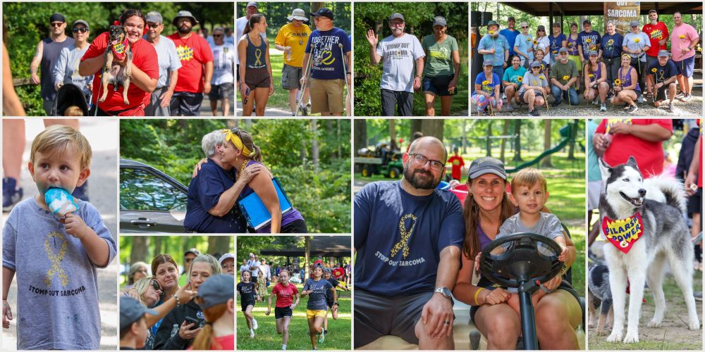
Support Stomp Out Sarcoma 2025!
I registered for the Stomp Out Sarcoma Fun Run/Walk in support of the @UMRogelCancerCenter! You can help make my run a success by making a donation. Your contribution will support Sarcoma Clinical Res...
michiganmedicine.donordrive.com
Kyle Perry
@kyledperrymd.bsky.social
· May 6
Kyle Perry
@kyledperrymd.bsky.social
· May 6
Kyle Perry
@kyledperrymd.bsky.social
· Apr 13
Kyle Perry
@kyledperrymd.bsky.social
· Apr 13
Kyle Perry
@kyledperrymd.bsky.social
· Apr 13
Kyle Perry
@kyledperrymd.bsky.social
· Mar 31
Kyle Perry
@kyledperrymd.bsky.social
· Mar 31
Kyle Perry
@kyledperrymd.bsky.social
· Mar 31
Kyle Perry
@kyledperrymd.bsky.social
· Mar 31
Kyle Perry
@kyledperrymd.bsky.social
· Mar 16
Kyle Perry
@kyledperrymd.bsky.social
· Mar 16
Kyle Perry
@kyledperrymd.bsky.social
· Mar 16
Kyle Perry
@kyledperrymd.bsky.social
· Feb 8
Kyle Perry
@kyledperrymd.bsky.social
· Feb 8