Louise Fets
@louisefets.bsky.social
620 followers
590 following
42 posts
Researcher with an interest in pharmacology and cancer metabolism, Group Leader @mrc-lms.bsky.social & CRUK Career Establishment Awardee
Posts
Media
Videos
Starter Packs
Pinned

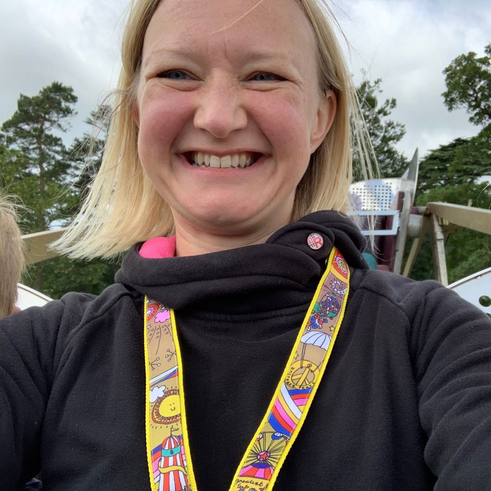
Louise Fets
@louisefets.bsky.social
· Jul 10
Louise Fets
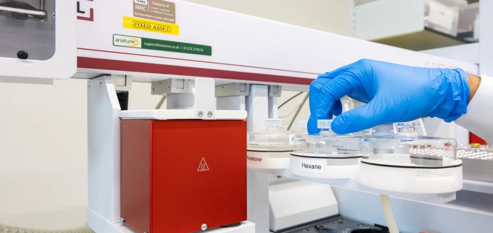
@louisefets.bsky.social
· Jul 7
Reposted by Louise Fets

Reposted by Louise Fets
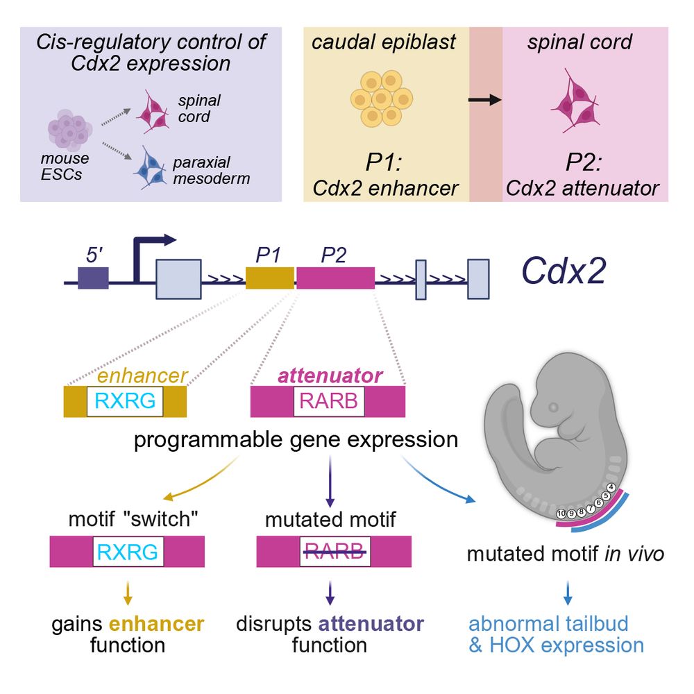
Louise Fets
@louisefets.bsky.social
· Jun 25
MRC Postdoctoral Research Scientist - MRC Laboratory of Medical Sciences
The MRC Laboratory of Medical Sciences (LMS) is a biomedical research institute where scientists and clinicians collaborate to advance the understanding of biology and its application to medicine. LMS...
lms.mrc.ac.uk
Louise Fets
@louisefets.bsky.social
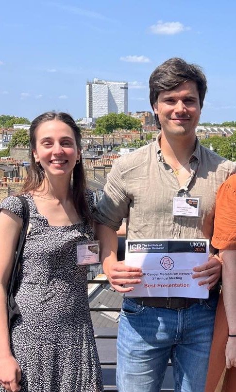
· Jun 14
Louise Fets
@louisefets.bsky.social
· Jun 12
Louise Fets
@louisefets.bsky.social
· Jun 12
Louise Fets
@louisefets.bsky.social
· Jun 12
Louise Fets
@louisefets.bsky.social
· Jun 12
Louise Fets
@louisefets.bsky.social
· Jun 12