epithelial mechanics fan club
@epimechfc.bsky.social
1.7K followers 220 following 750 posts
📚 We're your source for papers on various #EpithelialMechanics topics 🔍 & platform to share your research and passion 🧫🔬 Like to write a thread? Please DM us! 💬 @onenimesa.bsky.social & @juliaeckert.bsky.social 👉 https://epithelialmechanics.github.io
Posts Media Videos Starter Packs
Pinned
epimechfc.bsky.social
📚 Welcome to the Epithelial Mechanics Fan Club! 🌟

We're all about sharing research papers and threads. If you'd like to contribute, please send DM us! 💬

Our goal is to build a community that fosters knowledge sharing.

Explore our previous 🧵 here 👇
Reposted by epithelial mechanics fan club
epimechfc.bsky.social
1/ 🐣⚒️ To study mechanics, we need to apply prescribed forces - how can we do that in living tissues? I am @eigenp.bsky.social and in this thread I'll highlight our new approach for applying tension on epithelial tissues in vivo!
epimechfc.bsky.social
Thank you for reading the thread. I am @eigenp.bsky.social. For more follow @epimechfc.bsky.social bsky.app/profile/epim...
epimechfc.bsky.social
📚 Welcome to the Epithelial Mechanics Fan Club! 🌟

We're all about sharing research papers and threads. If you'd like to contribute, please send DM us! 💬

Our goal is to build a community that fosters knowledge sharing.

Explore our previous 🧵 here 👇
epimechfc.bsky.social
9/ Related reads: 🧪📖 🔬 🐣 📐
Harris et al. (2012, PNAS) bsky.app/profile/epim...
Charras & Yap (2018, Current Biology)
bsky.app/profile/epim...
Zhou, Kim & Davidson (2009, Development)
bsky.app/profile/epim...
epimechfc.bsky.social
Charras, G. and Yap, A. S. (2018). Tensile forces and mechanotransduction at cell–cell junctions. Current Biology, #EpithelialMechanicsReview www.cell.com/current-biol...
epimechfc.bsky.social
8/ Demo 3: Figure out the forces needed to unzip the neural tube (precursor to the brain!)
Morphogenetic force threshold, in vivo. ⚙️➡️🧠
epimechfc.bsky.social
7/ Demo 2: Disrupt actin in one region (🟢 = actin perturbed, 🔴 = wild-type) → test for non-cell-autonomous effects (mechanical cross-talk beyond the perturbed zone) 🧩
epimechfc.bsky.social
6/ You can do so much with it! Here are some case studies:
Demo 1: stretch the endoderm (ventral epithelium of the developing amniote 🐣 embryo, here in 🟢) → observe mechanical coupling with the subjacent mesoderm (brightfield ⬜) 🎥
epimechfc.bsky.social
5/ How it works:
🛠️ Two fine tungsten cantilevers pull on filter-paper tabs adhered to the epithelium.
📏 Cantilever bend → μN force readout (~1–100 μN).
The entire setup fits on a microscope stage, so you can image responses live. 🔬 bsky.app/profile/epim...
epimechfc.bsky.social
4/ Nonetheless, epithelial tissues in particular remain a challenge…
No need to FRET (😜) over it — we have a new tool that brings calibrated tensile control for epithelia in vivo!
epimechfc.bsky.social
3/ Luckily, @xaviertrepat.bsky.social lab wrote a comprehensive survey of methods 🧰 to measure/perturb stress - in vivo - including stretchers, deformable beads/droplets, laser ablation, and force inference
epimechfc.bsky.social
2/ Context: From monolayer tensile tests to junction mechanics and viscoelasticity @gcharras.bsky.social lab, we’ve learned loads in vitro. 🧫 📚 But in vivo? Harder
epimechfc.bsky.social
Khalilgharibi, N., Fouchard, J., Asadipour, N., ... & Charras, G. (2019). Stress relaxation in epithelial monolayers is controlled by the actomyosin cortex. Nature physics, 15(8), 839–847. doi.org/10.1038/s415... #EpithelialMechanics
epimechfc.bsky.social
1/ 🐣⚒️ To study mechanics, we need to apply prescribed forces - how can we do that in living tissues? I am @eigenp.bsky.social and in this thread I'll highlight our new approach for applying tension on epithelial tissues in vivo!
epimechfc.bsky.social
Zhou, J., Kim, H. Y., & Davidson, L. A. (2009). Actomyosin stiffens the vertebrate embryo during crucial stages of elongation and neural tube closure. #EpithelialMechanics doi.org/10.1242/dev....
epimechfc.bsky.social
Gómez-González, M., Latorre, E., Arroyo, M., & Trepat, X. (2020). Measuring mechanical stress in living tissues. Nature Reviews Physics, #EpithelialMechanics www.nature.com/articles/s42...
epimechfc.bsky.social
Charras, G. and Yap, A. S. (2018). Tensile forces and mechanotransduction at cell–cell junctions. Current Biology, #EpithelialMechanicsReview www.cell.com/current-biol...
epimechfc.bsky.social
Oikonomou, P., Calvary, L., Cirne, H. C., Welch, A. E., ... & Nerurkar, N. L. (2025). Application and measurement of tissue-scale tension in avian epithelia in vivo to study multiscale mechanics and inter-germ layer coupling. Development, #EpithelialMechanics doi.org/10.1242/dev....
epimechfc.bsky.social
Harris, A. R., Peter, L., Bellis, J., Baum, B., Kabla, A. J., & Charras, G. T. (2012). Characterizing the mechanics of cultured cell monolayers. Proceedings of the National Academy of Sciences of the United States of America, #EpithelialMechanics doi.org/10.1073/pnas...
epimechfc.bsky.social
Khalilgharibi, N., Fouchard, J., Asadipour, N., ... & Charras, G. (2019). Stress relaxation in epithelial monolayers is controlled by the actomyosin cortex. Nature physics, 15(8), 839–847. doi.org/10.1038/s415... #EpithelialMechanics
epimechfc.bsky.social
About symmetry-breaking in embryonic development
bsky.app/profile/epim...
epimechfc.bsky.social
How do dynamic forces influence symmetry-breaking in embryonic development?

Hi, I’m @dr-rajendra-s-negi.bsky.social & in this thread, I’d like to introduce some aspects of the interdisciplinary research on tissue dynamics.
epimechfc.bsky.social
This review explains "cell jamming," where cells stop moving due to crowding, tension, and reduced fluctuations, and suggests identifying the main cause in experiments doi.org/10.1016/j.ce...
epimechfc.bsky.social
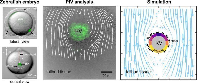
This research shows that organ movement through tissue generates viscoelastic drag forces, causing specific cell shape changes. These forces, observed in experimental studies, play a crucial role in shaping cells during development. doi.org/10.1016/j.cd...
epimechfc.bsky.social
This study reveals that asymmetric cell volume & shape changes in Kupffer's vesicle are regulated by ion flux, occurring independently of lumen expansion

Ion flux distinguishes intrinsic cellular mechanisms from lumen-generated forces in morphogenesis doi.org/10.7554/eLif...
epimechfc.bsky.social
Research suggests that fluid-like drag forces from an organ's movement through surrounding tissue can influence its structure and function.

This study examines zebrafish Kupffer's vesicle & the shape changes of its monociliate cells during development. doi.org/10.1016/j.bp...
epimechfc.bsky.social
Vertex & Voronoi models are used to study the mechanical properties & behaviors of cells and tissues.

Here the differences in boundary behavior in the 3D vertex and Voronoi models are presented: journals.plos.org/ploscompbiol...